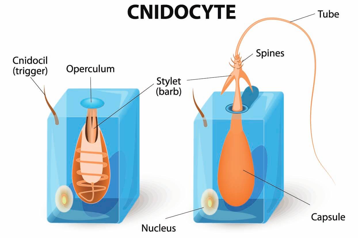
3d Reconstruction Of The Sea Anemones Stinging Organelle
3d Reconstruction Of The Sea A Video Eurekalert Science News Releases The stinging organelles of jellyfish, sea anemones, and other cnidarians, known as nematocysts, are remarkable cellular weapons used for both predation and defense. nematocysts consist of a. Serial images from electron microscopy were used to create a 3d reconstruction of the sea anemone's stinging organelle. credit: gibson lab, stowers institute for medical research.

Inside The Jellyfish S Sting Exploring The Micro Architecture Of A An ancient stinging organelle that evolved to deliver toxins could inspire the design of new medical delivery devices. during feeding, the sea anemone’s tentacles capture brine shrimp. video courtesy of the gibson lab, stowers institute for medical research. kansas city, mo—june 21, 2022—summertime beachgoers are all too familiar with the. Abstract. the stinging organelles of jellyfish, sea anemones, and other cnidarians, known as nematocysts, are remarkable cellular weapons used for both predation and defense. nematocysts consist of a pressurized capsule containing a coiled harpoon like thread. these structures are in turn built within specialized cells known as nematocytes. Here, using a combination of live and super resolution imaging, 3d electron microscopy and genetic perturbations, we define the step by step sequence of nematocyst operation in the model sea anemone nematostella vectensis. this analysis reveals the complex biomechanical transformations underpinning the operating mechanism of nematocysts, one of the nature’s most exquisite biological micro. Serial images from electron microscopy were used to create a 3d reconstruction of the sea anemone’s stinging organelle.

Polyp Cnidarian Coral Sea Anemone Britannica Here, using a combination of live and super resolution imaging, 3d electron microscopy and genetic perturbations, we define the step by step sequence of nematocyst operation in the model sea anemone nematostella vectensis. this analysis reveals the complex biomechanical transformations underpinning the operating mechanism of nematocysts, one of the nature’s most exquisite biological micro. Serial images from electron microscopy were used to create a 3d reconstruction of the sea anemone’s stinging organelle. The stinging organelle of the starlet sea anemone, nematostella vectensis. the study, published online in nature communications on june 17, 2022, was led by ahmet karabulut, a predoctoral. In sea 45 anemones, nematocyst capsules are sealed by three apical flaps connected to the stinging 46 thread22. this thread is composed of two distinct sub structures: a short, rigid and fibrous shaft 47 and a long thin tubule decorated with barbs16,18. the shaft is composed of three helically coiled.

Sea Anemone Facts And Beyond Biology Dictionary The stinging organelle of the starlet sea anemone, nematostella vectensis. the study, published online in nature communications on june 17, 2022, was led by ahmet karabulut, a predoctoral. In sea 45 anemones, nematocyst capsules are sealed by three apical flaps connected to the stinging 46 thread22. this thread is composed of two distinct sub structures: a short, rigid and fibrous shaft 47 and a long thin tubule decorated with barbs16,18. the shaft is composed of three helically coiled.

Diagram Of A Sea Anemone

Comments are closed.