A Labeled Diagram Of A Striated Muscle Striated Beef Muscle A Labeled

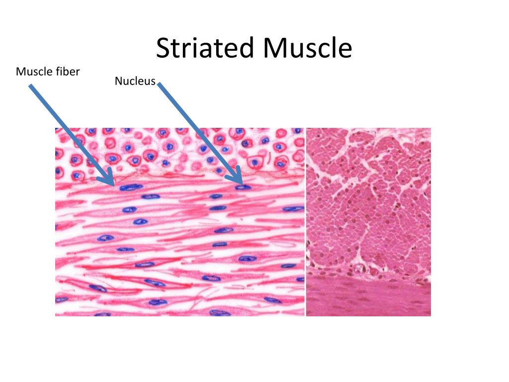
Diagram Of Striated Muscle Striated musculature. this type of tissue is found in skeletal muscles and is responsible for the voluntary movements of bones. striated musculature comprises of two types of tissues: skeletal muscle and cardiac muscle. skeletal muscle is the tissue that most muscles attached to bones are made of. hence the word "skeletal". Start studying striated (skeletal) muscle. learn vocabulary, terms, and more with flashcards, games, and other study tools. anatomy summer 2023 leg. 10 terms.

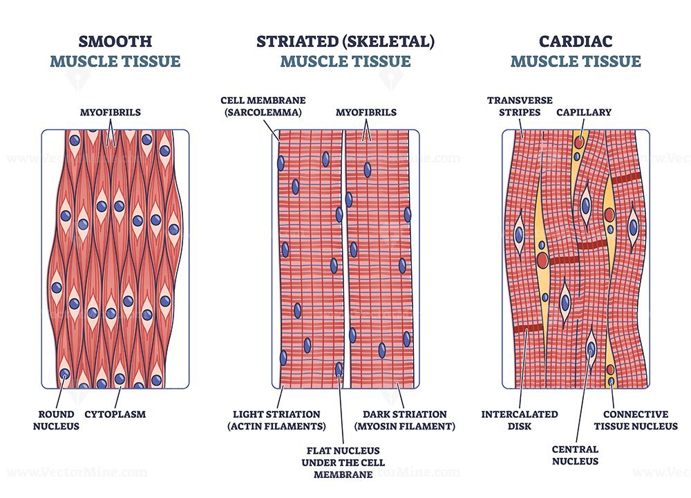
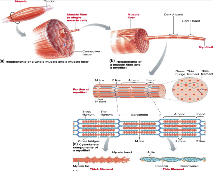
Ppt Striated Muscle Powerpoint Presentation Free Download Id 2513807 Fma. 67905. anatomical terminology. [edit on wikidata] striated muscle tissue is a muscle tissue that features repeating functional units called sarcomeres. the presence of sarcomeres manifests as a series of bands visible along the muscle fibers, which is responsible for the striated appearance observed in microscopic images of this tissue. Part of muscle fiber (cell) location. term. sarcalema. location. start studying label striated muscle. learn vocabulary, terms, and more with flashcards, games, and other study tools. Figure 10.2.2 – muscle fiber: a skeletal muscle fiber is surrounded by a plasma membrane called the sarcolemma, which contains sarcoplasm, the cytoplasm of muscle cells. a muscle fiber is composed of many myofibrils, which contain sarcomeres with light and dark regions that give the cell its striated appearance. the sarcomere. Cardiac muscle. cardiac muscle tissue, like skeletal muscle tissue, looks striated or striped. the bundles are branched, like a tree, but connected at both ends. unlike skeletal muscle tissue, the contraction of cardiac muscle tissue is usually not under conscious control, so it is called involuntary. smooth muscle.

Striated Muscles Definition Structure Types Functions Cbse Figure 10.2.2 – muscle fiber: a skeletal muscle fiber is surrounded by a plasma membrane called the sarcolemma, which contains sarcoplasm, the cytoplasm of muscle cells. a muscle fiber is composed of many myofibrils, which contain sarcomeres with light and dark regions that give the cell its striated appearance. the sarcomere. Cardiac muscle. cardiac muscle tissue, like skeletal muscle tissue, looks striated or striped. the bundles are branched, like a tree, but connected at both ends. unlike skeletal muscle tissue, the contraction of cardiac muscle tissue is usually not under conscious control, so it is called involuntary. smooth muscle. Figure 10.4 muscle fiber a skeletal muscle fiber is surrounded by a plasma membrane called the sarcolemma, which contains sarcoplasm, the cytoplasm of muscle cells. a muscle fiber is composed of many fibrils, which give the cell its striated appearance. Department of neurobiology and developmental sciences. university of arkansas for medical sciences. 4301 w. markham, slot 510. little rock, ar 72205. oct 9, 2014, 8:20 pm. this page focuses on illustrations and a study guide for striated skeletal muscle.

Comments are closed.