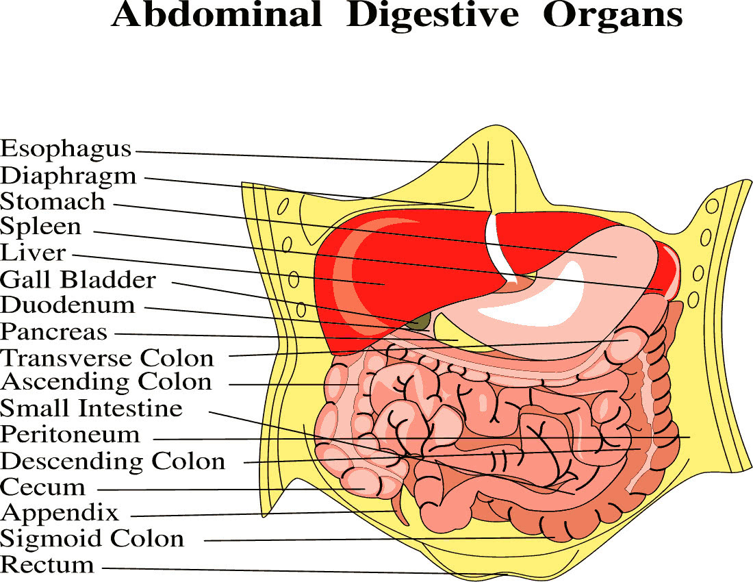
Abdominal Anatomy Abdominal Anatomy At University Of California San
Surface Anatomy Of The Abdomen The department of anatomy includes 19 faculty members with primary full time appointments, 15 faculty with joint appointments, and about 200 postdoctoral fellows and students engaged in research that covers questions in cell biology, developmental biology and the neurosciences. faculty have laboratories at both the parnassus heights campus. Benjamin yeh, md. professor. program director, body imaging fellowship. ph: (415) 353 3900. fax: (415) 353 7299. (415) 514 8888. the abdominal imaging experts at ucsf radiology diagnose and treat disorders of the liver, pancreas, colon, uterus, ovaries, prostate, and bladder with patient–centered imaging that uses the lowest levels of radiation.
Abdominal Anatomy Abdominal Anatomy At University Of California San After a needs assessment conducted by the department of surgery at the university of california, san francisco, school of medicine (ucsf) revealed a large interest in using surgical footage to enhance anatomy curricula, we set out to develop video tutorials using our extensive library of abdominal procedures, current video editing software, and. Abdominal ultrasound is a type of imaging test. it is used to look at organs in the abdomen, including the liver, gallbladder, spleen, pancreas, and kidneys. the blood vessels that lead to some of these organs, such as the inferior vena cava and aorta, can also be examined with ultrasound. At the university of california, san francisco school of medicine, the surgery and anatomy departments collaborated to create guided video tutorials using laparoscopic surgical footage to teach the anatomy of the lesser sac and gastroesophageal junction. methods: these tutorials are instructional adjuncts to a laparoscopy session on fresh. An abdominal magnetic resonance imaging scan is an imaging test that uses powerful magnets and radio waves. the waves create pictures of the inside of the belly area. it does not use radiation (x rays). single magnetic resonance imaging (mri) images are called slices. the images can be stored on a computer, viewed on a monitor, printed on film.

Human Anatomy Abdominal Cavity Diagram At the university of california, san francisco school of medicine, the surgery and anatomy departments collaborated to create guided video tutorials using laparoscopic surgical footage to teach the anatomy of the lesser sac and gastroesophageal junction. methods: these tutorials are instructional adjuncts to a laparoscopy session on fresh. An abdominal magnetic resonance imaging scan is an imaging test that uses powerful magnets and radio waves. the waves create pictures of the inside of the belly area. it does not use radiation (x rays). single magnetic resonance imaging (mri) images are called slices. the images can be stored on a computer, viewed on a monitor, printed on film. Benjamin m. yeh, md, is a professor in the abdominal imaging section in the department of radiology at the university of california, san francisco (ucsf). dr. yeh directs the nih funded contrast and ct research lab at ucsf which explores novel contrast enhanced imaging techniques for ct, dual energy ct and mri. A.d.a.m. student atlas of anatomy april 2008 19th august 2024: digital purchasing is currently unavailable on cambridge core. due to recent technical disruption affecting our publishing operation, we are experiencing some delays to publication.

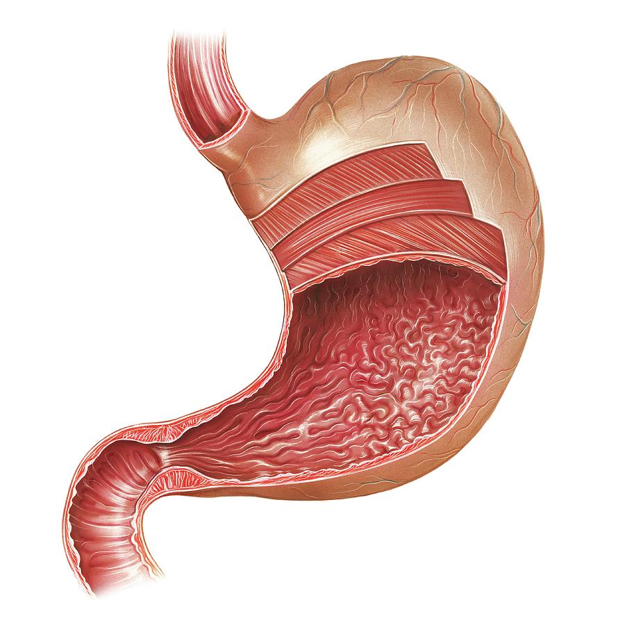
Stomach Cavity Anatomy Benjamin m. yeh, md, is a professor in the abdominal imaging section in the department of radiology at the university of california, san francisco (ucsf). dr. yeh directs the nih funded contrast and ct research lab at ucsf which explores novel contrast enhanced imaging techniques for ct, dual energy ct and mri. A.d.a.m. student atlas of anatomy april 2008 19th august 2024: digital purchasing is currently unavailable on cambridge core. due to recent technical disruption affecting our publishing operation, we are experiencing some delays to publication.

Abdominal Anatomy Medical Illustration Medivisuals

Comments are closed.