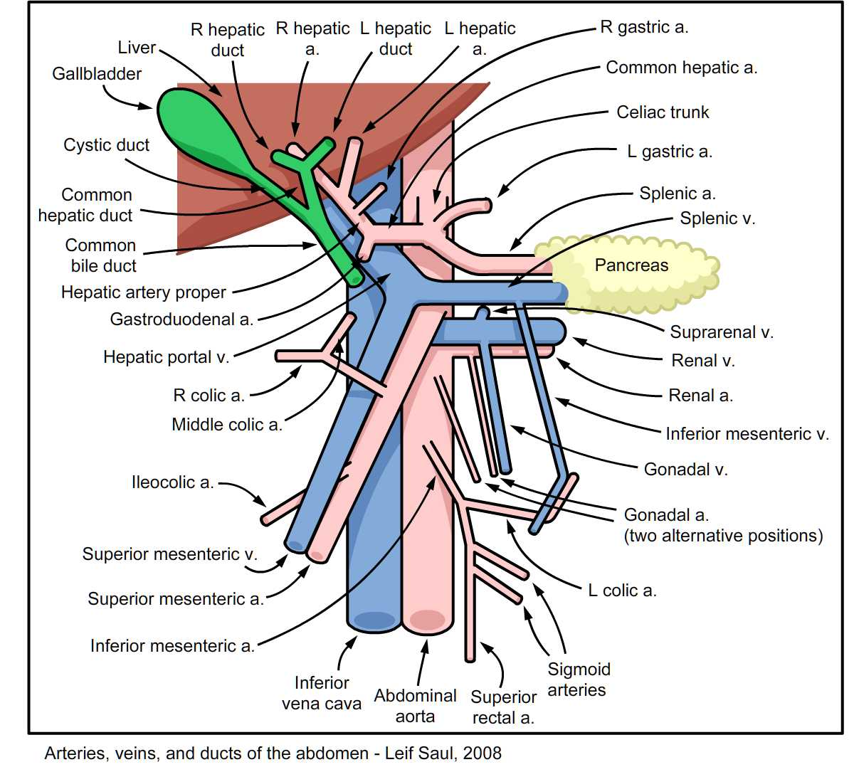
Abdominal Blood Vessels Labeled Arteries Veins Atlas Of Anatomy Hma

Abdominal Blood Vessels Labeled Arteries Veins Atlas Of Anatomy Hma The blood that goes out through those arteries comes back to the liver by way of the portal vein, as we’ll see in volume five of this atlas. the blood from the liver then returns to the main circulation through these large hepatic veins. the hepatic veins are very short. their number varies: here there are three. Here’s the abdominal aorta. it enters the abdomen by passing though the aortic opening in the diaphragm at the level of the twelfth thoracic vertebra. the aorta runs down on the front of the lumbar vertebral bodies, just to the left of the mid line. it ends by dividing at the level of l4, into the right and left common iliac arteries.

Anatomy Of The Abdominal Blood Vessels Stock Photo Al Vrogue Co As the abdomen and pelvis contain the majority of internal organs, these regions need to be supplied by an extensive network of arteries and veins. that being said, all arterial blood delivered to this region comes via branches of the abdominal aorta, and all venous blood eventually finds its way back to inferior vena cava (ivc). The portal vein is formed behind the pancreas. here's the pancreas. here's the duodenum; here it is again. here are the superior mesenteric, and inferior mesenteric veins, joining, and passing up behind the neck of the pancreas. to see more, we'll remove the left half of the pancreas. here behind the pancreas is the large splenic vein coming in. Checking the abdominal arteries. on a standard portal venous phase computed tomography (ct) series, you will get a balanced view of the vasculature that includes both major arteries and veins. on the other hand, an examination such as ct angiography obtains images earlier after contrast which allows you to see even small arterial branches. The abdominopelvic cavity is confined superiorly by the diaphragm and extends inferiorly into the pelvis. this cavity houses most digestive organs and parts of the urogenital system. the abdominopelvic vasculature is complex and supplies various organs and tissue layers across many planes. physiologic variation among vessels introduces diversity among patient populations. pathologies affecting.

Abdominal Arteries Anatomy Explore Organs Anatomy Diagram Checking the abdominal arteries. on a standard portal venous phase computed tomography (ct) series, you will get a balanced view of the vasculature that includes both major arteries and veins. on the other hand, an examination such as ct angiography obtains images earlier after contrast which allows you to see even small arterial branches. The abdominopelvic cavity is confined superiorly by the diaphragm and extends inferiorly into the pelvis. this cavity houses most digestive organs and parts of the urogenital system. the abdominopelvic vasculature is complex and supplies various organs and tissue layers across many planes. physiologic variation among vessels introduces diversity among patient populations. pathologies affecting. This website uses cookies to improve your experience while you navigate through the website. out of these, the cookies that are categorized as necessary are stored on your browser as they are essential for the working of basic functionalities of the website. Arteries transport blood away from the heart and branch into smaller vessels, forming arterioles. arterioles distribute blood to capillary beds, the sites of exchange with the body tissues. capillaries lead back to small vessels known as venules that flow into the larger veins and eventually back to the heart.

Comments are closed.