Advances In Eds Throughput From X Ray Counts To Solutions

Advances In Eds Throughput From X Ray Counts To Solutions Ongoing advancements in energy dispersive spectroscopy (eds) analysis are leading to higher count rates, better light element sensitivity, and improved energy resolution stability over a wide range of count rates. in this webinar we briefly review how the different parts of the eds system interact, from x rays leaving the sample to the production of useful data and where recent improvements. Ongoing advancements in energy dispersive spectroscopy (eds) analysis are leading to higher count rates, better light element sensitivity, and improved energ.

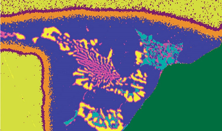
Advances In Eds Throughput From X Ray Counts To Solutions Youtube With the advances in energy dispersive spectrometry (eds) detectors, the technology has moved from single point and shoot spectrum collection to complete x ray imaging. in times past the output of the data collection was simply an image but modern systems have a full spectrum associated with each individual pixel in the image. The development of semiconductor based x ray detection in the 1960s led to the first successful energy dispersive x ray spectrometer (eds) using lithium compensated silicon [si(li) eds] operated on an electron column instrument, an epma . as compared to the narrow instantaneous energy range of the wds, the si(li) eds provided a view of the. This high throughput directly translates into greatly reduced times for x ray elemental mapping. operating in the x ray spectrum image (xsi) mode in which a complete eds spectrum is recorded at each beam position, sdd eds can capture maps of useful pixel density (256 ‗ 192) in less than 5 s that record information on major and minor constituents. Figure 5a shows the element maps of the galena sample plotted with “gross” x ray counts. in other words, with x ray counts that were not corrected after acquisition. also shown, for reference, is the wds element map for pb and for f. the elements identified as present by the eds spectrum are pb, o, f, s, ca, mn, cu, as, and sb.

A To Z Of Energy Dispersive X Ray Spectroscopy Eds Analyzetest This high throughput directly translates into greatly reduced times for x ray elemental mapping. operating in the x ray spectrum image (xsi) mode in which a complete eds spectrum is recorded at each beam position, sdd eds can capture maps of useful pixel density (256 ‗ 192) in less than 5 s that record information on major and minor constituents. Figure 5a shows the element maps of the galena sample plotted with “gross” x ray counts. in other words, with x ray counts that were not corrected after acquisition. also shown, for reference, is the wds element map for pb and for f. the elements identified as present by the eds spectrum are pb, o, f, s, ca, mn, cu, as, and sb. Electron excited x ray microanalysis performed in the scanning electron microscope with energy dispersive x ray spectrometry (eds) is a core technique for characterization of the microstructure of materials. the recent advances in eds performance with the silicon drift detector (sdd) enable accuracy and precision equivalent to that of the high spectral resolution wavelength dispersive. Recording adequate counts in the spectrum: the confidence of peak identification in qualitative analysis and the precision of quantitative eds x ray microanalysis benefit by increasing the number of measured x ray counts recorded in the spectrum. what constitutes an adequate number of x ray counts depends on the particular analytical task.

Comments are closed.