Amorphous Urate Crystals Of Urine Sediment Under Microscopy Medical

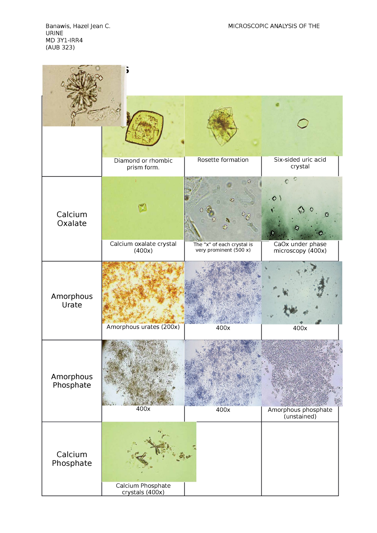
Amorphous Urates In Urine Crystals in urine: types, causes, symptoms & treatment. Urinary crystal formation is a marker of urinary supersaturation with various substances. crystals can occur secondary to inherited diseases, metabolic disorders, and medications. the presence of crystals in the urine is not always pathognomic for an abnormal metabolic or renal condition, as uric acid, calcium oxalate, calcium phosphate (cap), and drug induced crystals can also be observed.

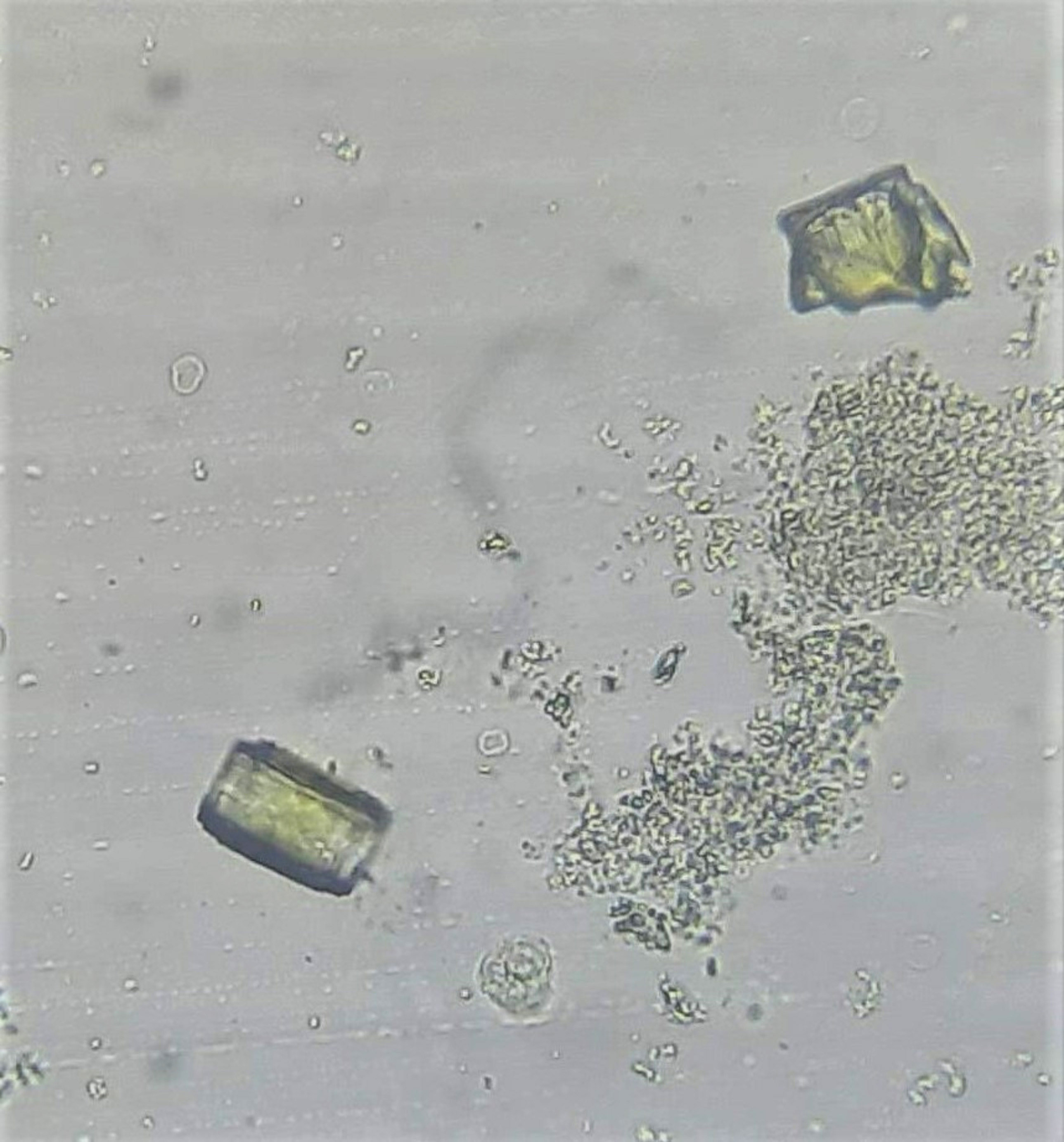
Amorphous Urate Crystals Of Urine Sediment Under Microscopyођ Amorphous urate crystals should be suspected when a turbid urine has a slightly pink appearance, a high specific gravity, and an acid ph. the sediment is pink because of the adherence of uroerythrin. the color is often referred to as “brick dust.” 1 , 2 , 5 amorphous urates will rapidly dissolve when urine specimens are warmed to 60°c. Differential identification of urine crystals with morphologic. Automated urine technology and centralized laboratory testing are becoming the standard for providing urinalysis data to clinicians, including nephrologists. this trend has had the unintended consequence of making examination of urine sediment by nephrologists a relatively rare event. in addition, the nephrology community appears to have lost interest in and forgotten the utility of provider. Ment to 10 μl of 0.9% saline, making a 1:2 di lution. the presence of amorphous. urates was graded as 0, 1 , or 2 as shown in figure 2. we treated 10 μl of urine sediment with 10 μl of 50 mm.

Amorphous Urates In Urine Automated urine technology and centralized laboratory testing are becoming the standard for providing urinalysis data to clinicians, including nephrologists. this trend has had the unintended consequence of making examination of urine sediment by nephrologists a relatively rare event. in addition, the nephrology community appears to have lost interest in and forgotten the utility of provider. Ment to 10 μl of 0.9% saline, making a 1:2 di lution. the presence of amorphous. urates was graded as 0, 1 , or 2 as shown in figure 2. we treated 10 μl of urine sediment with 10 μl of 50 mm. Prewarming unspun specimens to 60°c for 90 seconds dissolved most amorphous urates. conclusion: the protocol to eliminate amorphous urate crystals is to prewarm the specimen before testing. adding 50 mm naoh to sediment dissolves amorphous urates to enhance the visibility of bacteria and yeast but has a deleterious effect on wbc and rbc. Shown in figure 2. we treated 10 μl of urine sediment with 10 μl of. 50 mm naoh for a 1:2 dilution; 5 μl of urine sediment was treated with. 15 μl of 50 mm naoh for a 1:4 dilution. e presence.

Amorphous Urates In Urine Prewarming unspun specimens to 60°c for 90 seconds dissolved most amorphous urates. conclusion: the protocol to eliminate amorphous urate crystals is to prewarm the specimen before testing. adding 50 mm naoh to sediment dissolves amorphous urates to enhance the visibility of bacteria and yeast but has a deleterious effect on wbc and rbc. Shown in figure 2. we treated 10 μl of urine sediment with 10 μl of. 50 mm naoh for a 1:2 dilution; 5 μl of urine sediment was treated with. 15 μl of 50 mm naoh for a 1:4 dilution. e presence.

Comments are closed.