Anatomy Of The Cervical Spine Region Showing Neck Ver Vrogue Co

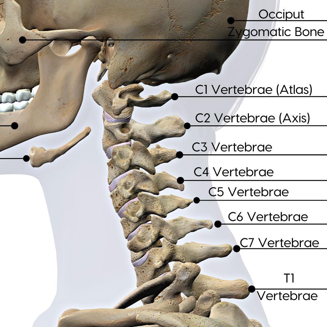
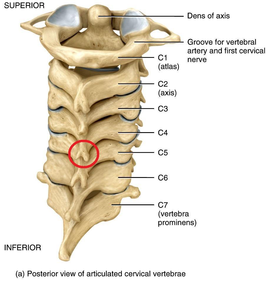
Cervical Spine With Ligaments And Nerves Posterior Vi Vrogue Co The cervical spine is the most superior portion of the vertebral column, lying between the cranium and the thoracic vertebrae. it consists of seven distinct vertebrae, two of which are given unique names: the first cervical vertebrae (c1) is known as the atlas. the second cervical vertebrae (c2) is known as the axis. Explore the anatomy and function of the cervical vertebrae with innerbody's interactive 3d model. the cervical vertebrae of the spine consist of seven bony rings that reside in the neck between the base of the skull and the thoracic vertebrae in the trunk. among the vertebrae of the spinal column, the cervical vertebrae are the thinnest and.

Anatomy Of The Cervical Spine Region Showing Neck Ver Vrogue Co Cervical spondylosis, also called arthritis of the neck, is the age related slow degeneration of your disks and joints in your cervical spine. cervical spinal cord injury. a cervical spinal cord injury is an injury to your cervical vertebrae. most spinal cord injuries are the result of a sudden, traumatic blow to the vertebrae. They form a natural inward curvature, sometimes called a lordotic curve. the upper cervical vertebrae are smaller and more mobile, while the lower ones are bigger to handle heavier loads from the neck and head. c3 through c6 are considered typical vertebrae because they share the same basic features with most of the spine’s vertebrae. Functions. support the head’s weight and allow a wide range of head and neck movements, like nodding and rotation. protect the spinal cord as it passes through the hollow space encircled by the 7 bones. the c1 to c6 vertebrae contain small holes that allow the vertebral artery, vein, and sympathetic nerves to pass through and carry blood to. Synonyms: vertebrae c1 c7. the cervical portion of the spine is an important one anatomically and clinically. it is within this region that the nerves to the arms arise via the brachial plexus, and where the cervical plexus forms providing innervation to the diaphragm among other structures. the cervical spine also allows passage of important.

Labeled Diagram Of Cervical Spine Vrogue Co Functions. support the head’s weight and allow a wide range of head and neck movements, like nodding and rotation. protect the spinal cord as it passes through the hollow space encircled by the 7 bones. the c1 to c6 vertebrae contain small holes that allow the vertebral artery, vein, and sympathetic nerves to pass through and carry blood to. Synonyms: vertebrae c1 c7. the cervical portion of the spine is an important one anatomically and clinically. it is within this region that the nerves to the arms arise via the brachial plexus, and where the cervical plexus forms providing innervation to the diaphragm among other structures. the cervical spine also allows passage of important. The cervical spine comprises 7 vertebrae (c1 to c7) and is divided into 2 major segments. the 2 most cephalad vertebrae, the atlas (c1) and the axis (c2), form the craniocervical junction (ccj) together with the occiput. the 5 cervical vertebrae caudad, c3 to c7, comprise the subaxial spine and are referred to by number (see image. C2 c3 joint. participates is subaxial (c2 c7) cervical motion that provides. 50° of cervical spine flexion extension. 50° of cervical spine rotation. 60° of lateral bending. c2 blood supply. a vascular watershed exists between the apex and the base of the odontoid. apex is supplied by branches of the internal carotid artery.

Cervical Spine Radiographic Anatomy Radiologypics Com Vrogue Co The cervical spine comprises 7 vertebrae (c1 to c7) and is divided into 2 major segments. the 2 most cephalad vertebrae, the atlas (c1) and the axis (c2), form the craniocervical junction (ccj) together with the occiput. the 5 cervical vertebrae caudad, c3 to c7, comprise the subaxial spine and are referred to by number (see image. C2 c3 joint. participates is subaxial (c2 c7) cervical motion that provides. 50° of cervical spine flexion extension. 50° of cervical spine rotation. 60° of lateral bending. c2 blood supply. a vascular watershed exists between the apex and the base of the odontoid. apex is supplied by branches of the internal carotid artery.

Comments are closed.