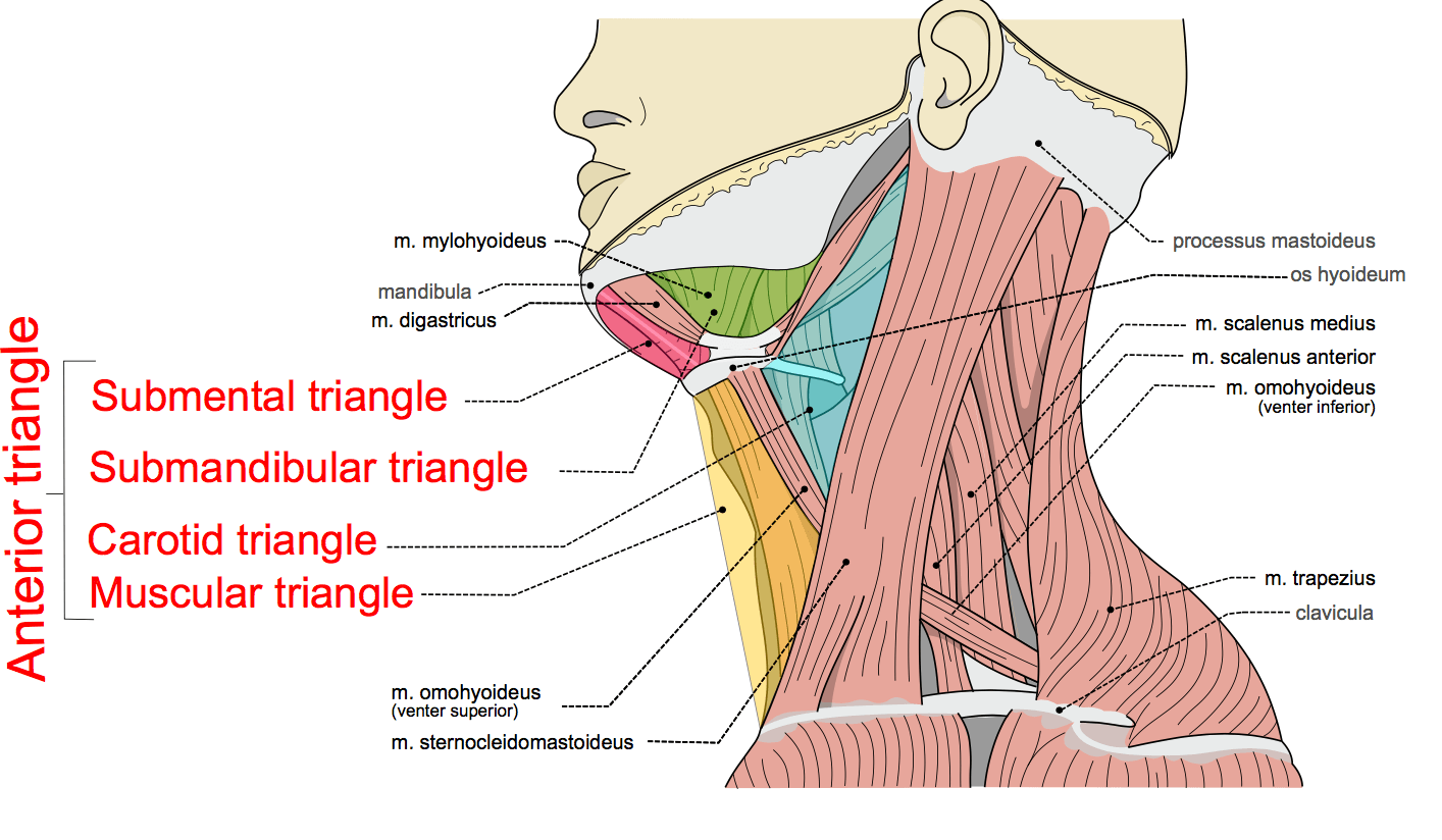
Anterior Triangle Of The Neck Boundaries Subdivisions Structures Anatomy Animated

Anterior Triangle Of The Neck Boundaries Subdivisions Structures Unlock the secrets of the neck's anterior triangle! dive into the crucial anatomy that governs respiration, swallowing, and speech, anchored by structures li. The submental triangle in the neck is situated underneath the chin. it contains the submental lymph nodes, which filter lymph draining from the floor of the mouth and parts of the tongue. it is bounded: inferiorly – hyoid bone. medially – midline of the neck. laterally – anterior belly of the digastric.

Triangles Of The Neck Part 1 The Anterior Triangle Medical Exam Prep The s ternocleidomastoid muscle divides the neck into the two major neck triangles; the anterior triangle and the posterior triangle of the neck, each of them containing a few subdivisions. the triangles of the neck are important because of their contents, as they house all the neck structures, including glands, nerves, vessels and lymph nodes. Boundariesmedial: the anterior median plane of the necklateral: sternocleidomastoidsuperior: base of the mandible and a line joining the angle of the mandibl. The neck is divided in two major triangles: anterior and posterior, based mainly on the borders of the sternocleidomastoid, or scm, and trapezius muscles, as well as other muscular and bony structures found in the neck. these regions provide a clear location regarding the structures, injuries or pathologies involving the neck. The limits of the neck are: medial: midline of the neck. lateral: anterior margin of trapezius. superior: inferior border of the mandible. inferior: superior border of the clavicle. the neck can further be divided into the anterior triangle and the posterior triangle. the muscle which delineates these two regions is the sternocleidomastoid (scm).

Anterior Triangle Of Neck Anatomy The neck is divided in two major triangles: anterior and posterior, based mainly on the borders of the sternocleidomastoid, or scm, and trapezius muscles, as well as other muscular and bony structures found in the neck. these regions provide a clear location regarding the structures, injuries or pathologies involving the neck. The limits of the neck are: medial: midline of the neck. lateral: anterior margin of trapezius. superior: inferior border of the mandible. inferior: superior border of the clavicle. the neck can further be divided into the anterior triangle and the posterior triangle. the muscle which delineates these two regions is the sternocleidomastoid (scm). Anterior belly: mylohyoid nerve, from mandibular division of trigeminal nerve (v); posterior belly: facial nerve (vii) forms two sides of the submandibular triangle. stylohyoid (tg7 21, tg7 34) posterior border of styloid process. splits around intermediate tendon of digastric to insert on the body of the hyoid bone. The boundaries of muscular triangle are as follows: anteriorly: anterior median line of the neck, extending from hyoid bone to the suprasternal notch. posterosuperiorly: superior belly of the omohyoid. posteroinferior: anterior border of sternocleidomastoid. floor is formed by sternothyroid, sternohyoid and thyrohyoid muscles.

Comments are closed.