Biol 230 Lab Manual Streptococcus Pneumoniae On Blood Agar

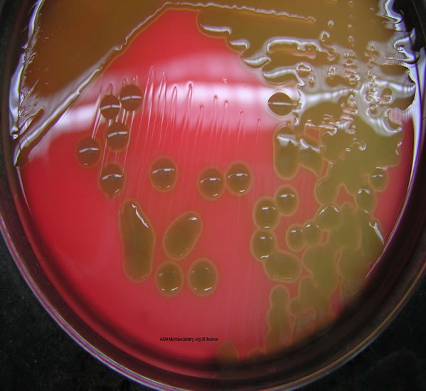
Biol 230 Lab Manual The Pneumococcus On Blood Agar Biol 230 lab manual: streptococcus pneumoniae on blood agar. note the mucoid colonies, alpha hemolysis (greenish discolorization of the red blood cells around the colonies) and sensitivity to the drug optochin in the taxo p® disc. photograph from from microbelibrary.org. courtesy of rebecca buxton, university of utah. Streptococcus pneumoniae. note the mucoid, transluscent colonies and the alpha hemolysis (partial hemolysis typically accompanied by a greenish discolorization of the agar around and under the growth). photograph from microbelibrary.org. courtesy of rebecca buxton, university of utah.

16 Streptococcus Pneumoniae Growing On Blood Agar Download Fig. 12: streptococcus pneumoniae growing on blood agar with a taxo p disk (indirect lighting) note the mucoid colonies, alpha hemolysis (greenish discolorization of the red blood cells around the colonies) and sensitivity to the drug optochin in the taxo p® disk. photograph from from microbelibrary.org. Culture and sensitivity inoculate sample onto blood agar and chocolate agar plate. incubate at 37°c with 5 10% co 2 for 24 – 48 hours.; colony morphology. colonies on blood agar plate are small (0.5 mm), round, translucent, or mucoid with alpha hemolysis (a green discoloration of the agar around the colonies). Other important pore forming membrane disrupting toxins include alpha toxin of staphylococcus aureus and pneumolysin of streptococcus pneumoniae. (openstax cnx, 2018) (openstax cnx, 2018) blood agar (tsb w 5%sb) pancreatic digest of casein 14.5 g l peptic digest of soybean meal 5.0 g l sodium chloride 5.0 g l, agar 14.0 g l, defibrinated sheep blood 5.0%. Figure \(\pageindex{1}\): streptococcus pneumoniae growing on blood agar with a taxo p disk (indirect lighting). note the mucoid colonies, alpha hemolysis (greenish discolorization of the red blood cells around the colonies) and sensitivity to the drug optochin in the taxo p® disk. (courtesy of rebecca buxton, university of utah) 3. bile.

Comments are closed.