Cardiac Muscle And Electrical Activity Anatomy And Physiology I

Cardiac Muscle And Electrical Activity в Anatomy And Physiology Action potential in cardiac contractile cells. (a) note the long plateau phase due to the influx of calcium ions. the extended refractory period allows the cell to fully contract before another electrical event can occur. (b) the action potential for heart muscle is compared to that of skeletal muscle. Figure 19.4.1 – major factors influencing cardiac output: cardiac output is influenced by heart rate and stroke volume, both of which are also variable. svs are also used to calculate ejection fraction, which is the portion of the blood that is pumped or ejected from the heart with each contraction.

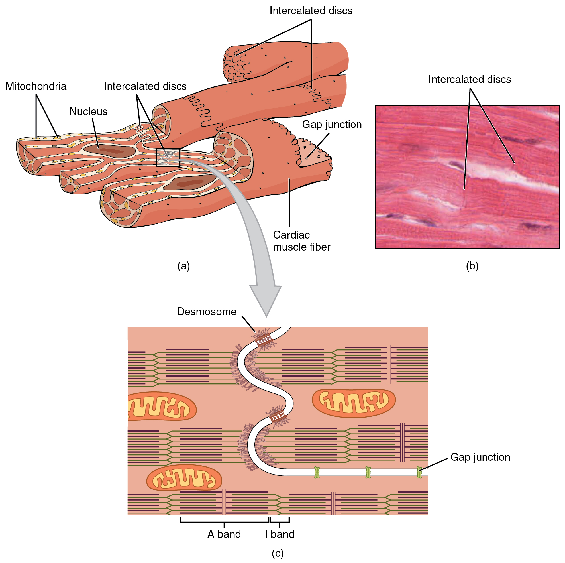
Cardiac Muscle And Electrical Activity в Anatomy And Physiology Introduction. cardiac muscle also called the myocardium, is one of three major categories of muscles found within the human body, along with smooth muscle and skeletal muscle. cardiac muscle, like skeletal muscle, is made up of sarcomeres that allow for contractility. however, unlike skeletal muscle, cardiac muscle is under involuntary control. Cardiac muscle tissue is only found in the heart. highly coordinated contractions of cardiac muscle pump blood into the vessels of the circulatory system. similar to skeletal muscle, cardiac muscle is striated and organized into sarcomeres, possessing the same banding organization as skeletal muscle (figure 10.21). however, cardiac muscle. There are two major types of cardiac muscle cells: myocardial contractile cells and myocardial conducting cells. the myocardial contractile cells constitute the bulk (99 percent) of the cells in the atria and ventricles. contractile cells conduct impulses and are responsible for contractions that pump blood through the body. The t wave (and a portion of the qrs complex) measure the electrical activity associated with repolarization, the physiological reset required to prepare for the next cardiac cycle. figure 17.3.5 17.3. 5: electrocardiogram. a normal tracing shows the p wave, qrs complex, and t wave. also indicated are the pr, qt, qrs, and st intervals, plus the.

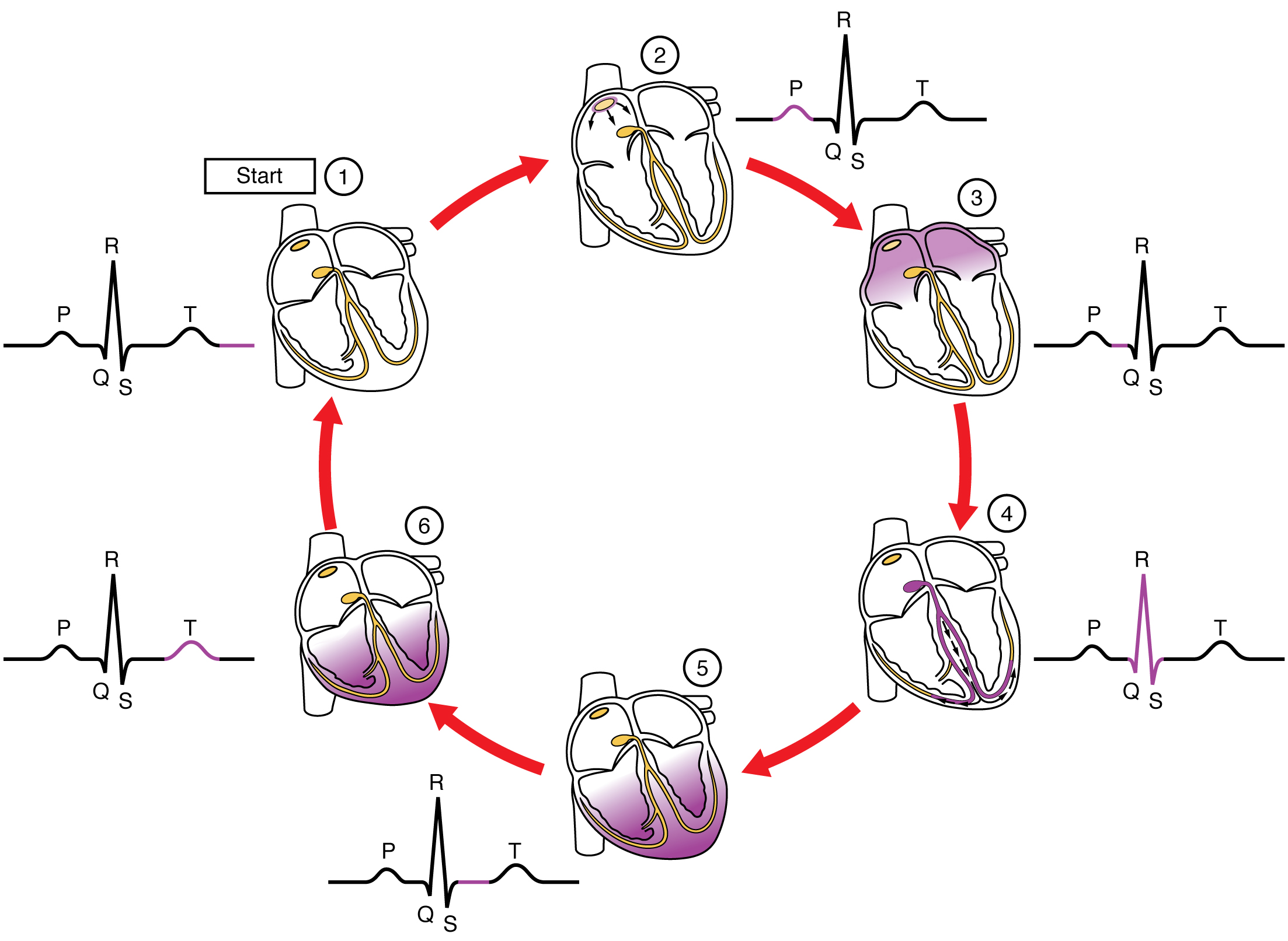
The Heart And Ekg I Anatomy Of The Heart 1 Heart Valves There are two major types of cardiac muscle cells: myocardial contractile cells and myocardial conducting cells. the myocardial contractile cells constitute the bulk (99 percent) of the cells in the atria and ventricles. contractile cells conduct impulses and are responsible for contractions that pump blood through the body. The t wave (and a portion of the qrs complex) measure the electrical activity associated with repolarization, the physiological reset required to prepare for the next cardiac cycle. figure 17.3.5 17.3. 5: electrocardiogram. a normal tracing shows the p wave, qrs complex, and t wave. also indicated are the pr, qt, qrs, and st intervals, plus the. Figure 17.3.4 17.3. 4 show the spread of the electrical wave through these structures and the myocardial contractile cells. figure 17.3.4 17.3. 4: cardiac conduction. (1) the sinoatrial (sa) node and the remainder of the conducting system are at rest. (2) the sa node initiates the electrical wave, which sweeps across the atria. The cardiac cycle includes two parts, called diastole and systole, which are illustrated in figure 10.4.1 10.4. 1. during diastole, the atria contract and pump blood into the ventricles, while the ventricles relax and fill with blood from the atria. during systole, the atria relax and collect blood from the lungs and body, while the ventricles.

19 2 Cardiac Muscle And Electrical Activity Anatomy And Physiologyо Figure 17.3.4 17.3. 4 show the spread of the electrical wave through these structures and the myocardial contractile cells. figure 17.3.4 17.3. 4: cardiac conduction. (1) the sinoatrial (sa) node and the remainder of the conducting system are at rest. (2) the sa node initiates the electrical wave, which sweeps across the atria. The cardiac cycle includes two parts, called diastole and systole, which are illustrated in figure 10.4.1 10.4. 1. during diastole, the atria contract and pump blood into the ventricles, while the ventricles relax and fill with blood from the atria. during systole, the atria relax and collect blood from the lungs and body, while the ventricles.

Heart Anatomy Electrical Pathway

Comments are closed.