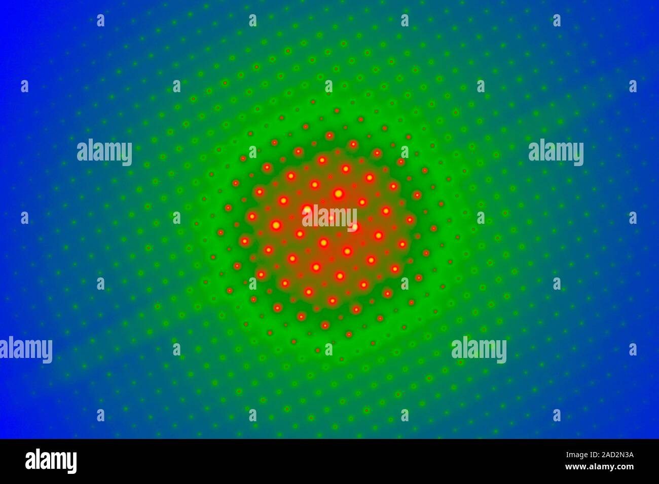
Crystallography Electron Diffraction Pattern Of Molybdenum

Crystallography Electron Diffraction Pattern Of Molybdenum Youtube Construction and analysis of an electron diffraction pattern of molybdenum, which has a body centred cubic crystal structure. construction of a twin electron. Powder x ray diffraction (xrd) was developed in 1916 by debye (figure 7.3.12) and scherrer (figure 7.3.13) as a technique that could be applied where traditional single crystal diffraction cannot be performed. this includes cases where the sample cannot be prepared as a single crystal of sufficient size and quality.

Coloured Transmission Electron Micrograph Tem Of The Electron Monochromators and filters are used to produce monochromatic x ray light. this narrow wavelength range is essential for diffraction calculations. for instance, a zirconium filter can be used to cut out unwanted wavelengths from a molybdenum metal target (see figure 4). the molybdenum target will produce x rays with two wavelengths. An electron‐diffraction pattern contains two kinds of information about a crystal. the positions of the diffraction spots are related to the unit‐cell parameters and the lattice type (see section 2.6). Proteins can also form crystals. • under certain conditions, entire proteins will pack into a regular grid (a lattice) multiple views of the crystal formed by an immunoglobulin binding domain (pdb entry 1pgb) note: the protein forms a regular pattern as with the able salt crystal. there’s a lot of “open space” not filled by the protein. Beam electron diffraction (cbed), in which a focused electron probe beam is used to obtain diffraction patterns from regions as small as 10 a. both tech niques provide a two dimensional pattern of diffraction spots, which can be highly symmetrical when a single crystal is oriented precisely along a crystal lographic direction.

Electron Diffraction Patterns Of Molybdenum Carbide Prepared With A Proteins can also form crystals. • under certain conditions, entire proteins will pack into a regular grid (a lattice) multiple views of the crystal formed by an immunoglobulin binding domain (pdb entry 1pgb) note: the protein forms a regular pattern as with the able salt crystal. there’s a lot of “open space” not filled by the protein. Beam electron diffraction (cbed), in which a focused electron probe beam is used to obtain diffraction patterns from regions as small as 10 a. both tech niques provide a two dimensional pattern of diffraction spots, which can be highly symmetrical when a single crystal is oriented precisely along a crystal lographic direction. Chapter 9 illustrates the intensities of x ray diffracted beams and considers atomic scattering factors, the structure factor equation, and its applications. next, it analyses the broadening of diffracted beams and the scherrer equation. the chapter examines the concept of integrated intensity; crystal size and perfection via mosaic structure. 2. x rays are electromagnetic waves. 3. the wavelength of the x rays is of the same order of magnitude (1 to 3) as the distance repeated by the motifs (ions, atoms, molecules, or assemblies thereof) in the crystals. x ray diffraction is a particular case of consistent radiation scattering. 1.

Comments are closed.