Deep Neck Spaces And Deep Cervical Fascia Anatomy Radio

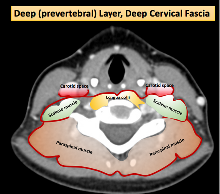
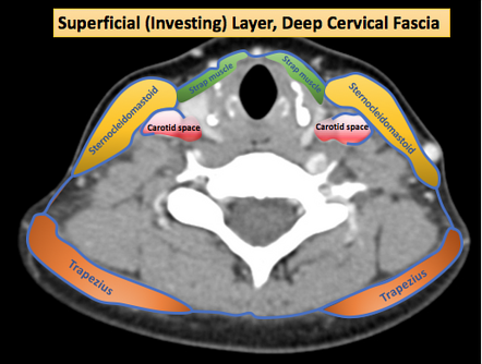
Deep Layer Of The Deep Cervical Fascia Radiology Reference Article Deep spaces of the head and neck. The spaces approach to the head and neck is based on compartments defined by the layers of the deep cervical fascia. each space contains unique contents which are subject to a predictable set of disease processes. localization of lesions to a particular space allows the generation of a limited radiologic differential diagnosis.

Infrahyoid Neck Spaces Defined By The Deep Cervical Fascia With Anatomy, head and neck, deep cervical neck fascia. Deep spaces of the head and neck annotated mri. The neck has 2 layers of fascia, the superficial and deep layers. the superficial cervical fascia is a thin layer that consists mainly of loose areolar connective tissue and adipose tissue that extends from the head to the thorax and the shoulders to the axilla. 1 it lies between the dermis of the skin and the deep cervical fascia and contains the platysma, muscles of facial expression. Fig. 4.1. fascial layers of the neck. (a) 3d illustration neck anatomy: deep cervical fascial layers defining the deep neck space in the supra and infra hyoid head and neck: investing layer (red), visceral layer (blue) and perivertebral or deep layer (orange). in part, these define a series of columns allowing craniocaudal spread of disease.

Imaging Anatomy Of Deep Neck Spaces Otolaryngologic Clinics Of North The neck has 2 layers of fascia, the superficial and deep layers. the superficial cervical fascia is a thin layer that consists mainly of loose areolar connective tissue and adipose tissue that extends from the head to the thorax and the shoulders to the axilla. 1 it lies between the dermis of the skin and the deep cervical fascia and contains the platysma, muscles of facial expression. Fig. 4.1. fascial layers of the neck. (a) 3d illustration neck anatomy: deep cervical fascial layers defining the deep neck space in the supra and infra hyoid head and neck: investing layer (red), visceral layer (blue) and perivertebral or deep layer (orange). in part, these define a series of columns allowing craniocaudal spread of disease. Understanding the deep neck anatomy is crucial for predicting the spread of infection and guiding treatment strategies. the superficial and deep cervical fascial planes form a series of compartments and spaces including the retropharyngeal, danger, prevertebral, carotid, parapharyngeal, submandibular, sublingual, parotid, masticator, temporal, and infrahyoid spaces. Head and neck imaging is an intimidating subject for many radiologists because of the complex anatomy and potentially serious consequences of delayed or improper diagnosis of the diverse abnormalities involving this region. the purpose of this article is to help radiologists to understand the intricate anatomy of the head and neck and to review the imaging appearances of a variety of.

Deep Neck Spaces And Deep Cervical Fascia Anatomy Radiolog Understanding the deep neck anatomy is crucial for predicting the spread of infection and guiding treatment strategies. the superficial and deep cervical fascial planes form a series of compartments and spaces including the retropharyngeal, danger, prevertebral, carotid, parapharyngeal, submandibular, sublingual, parotid, masticator, temporal, and infrahyoid spaces. Head and neck imaging is an intimidating subject for many radiologists because of the complex anatomy and potentially serious consequences of delayed or improper diagnosis of the diverse abnormalities involving this region. the purpose of this article is to help radiologists to understand the intricate anatomy of the head and neck and to review the imaging appearances of a variety of.

Deep Cervical Fascia Diagram Image Radiopaedia Org

Comments are closed.