Dermatomes Myotomes Dermatome By Theraspot Medium Dermatomes Chart

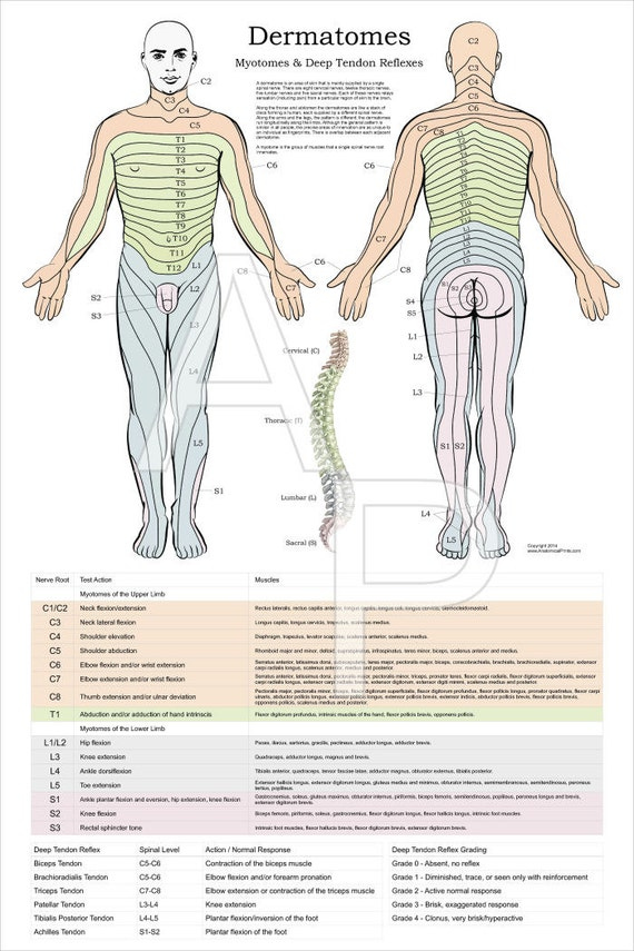
Dermatomes Myotomes Dermatome By Theraspot Medium Dermatomes Chart Dermatomes & myotomes. a dermatome is an area of skin supplied by sensory neurons that arise from a spinal nerve ganglion. it is an area of skin that is mainly supplied by afferent nerve fibers. A dermatome is an area of skin supplied by a single spinal nerve. there are 31 pairs of spinal nerves, forming nerve roots that branch from your spinal cord, but only 30 dermatomes. your spinal.

Dermatomes And Myotomes Chart Pdf Dermatome Map Dermatomes are areas of skin on your body that rely on specific nerve connections on your spine. in this way, dermatomes are much like a map. the nature of that connection means that dermatomes can help a healthcare provider detect and diagnose conditions or problems affecting your spine, spinal cord or spinal nerves. The term “ dermatome ” is a combination of two greek words; “derma” meaning “skin”, and “tome”, meaning “cutting” or “thin segment”. dermatomes are areas of the skin whose sensory distribution is innervated by the afferent nerve fibres from the dorsal root of a specific single spinal nerve root, which is that portion of. The lumbar plexus dermatomes and myotomes frequently play an essential function in finding out where the harm is originating from, providing doctors a hint regarding where to look for indications of infection, swelling, or injury. common diseases that might be partly determined through the dermatome chart consist of: spinal injury (from a fall. Dermatomes. a dermatome is an area of skin supplied by a single spinal nerve. if you imagine the human body as a map, each dermatome represents the area of skin supplied with sensation by a specific nerve root. it is important to bear in mind that the dermatomes of the head are supplied by branches v1, v2 and v3 of the trigeminal nerve.

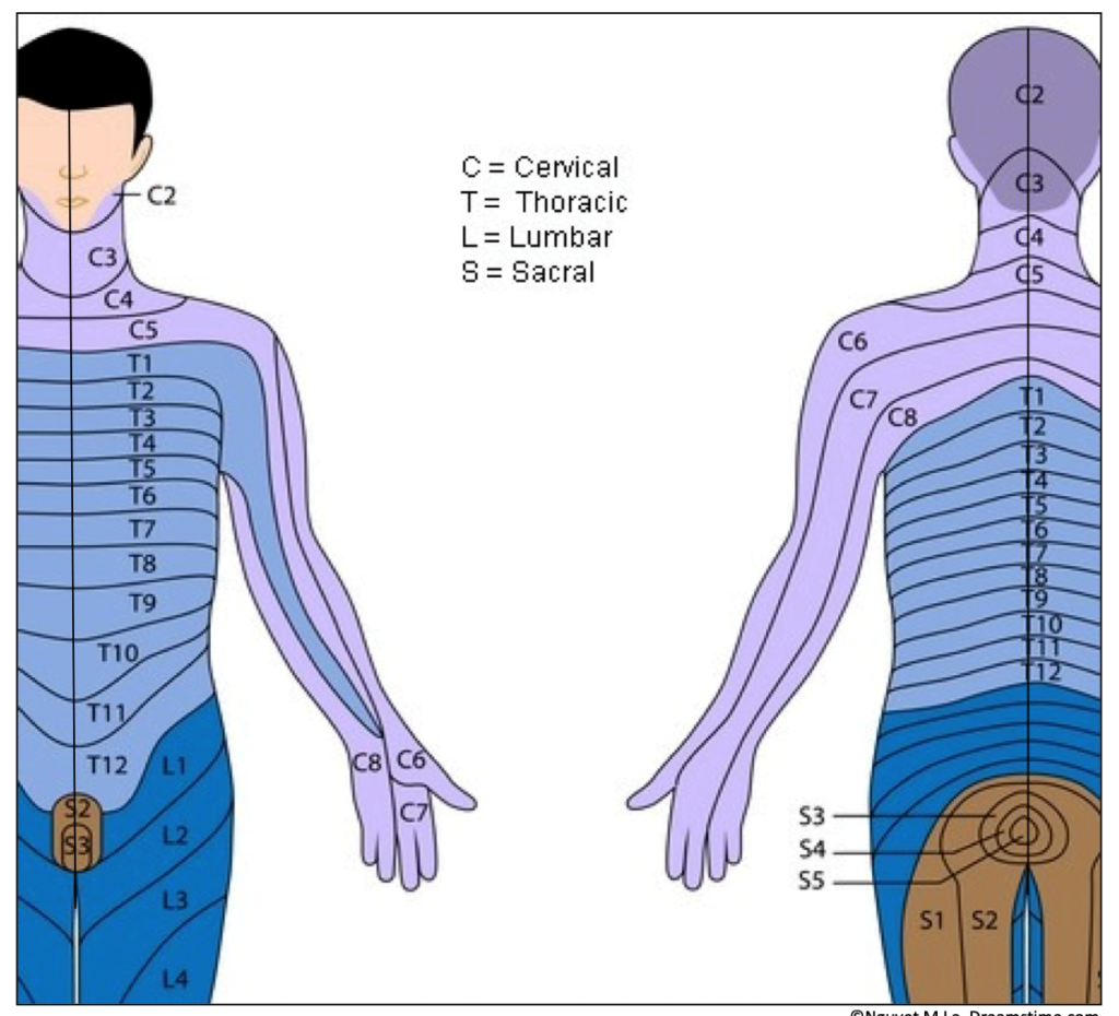
Lower Extremity Dermatomes And Myotomes Medical Anatomy Medical The lumbar plexus dermatomes and myotomes frequently play an essential function in finding out where the harm is originating from, providing doctors a hint regarding where to look for indications of infection, swelling, or injury. common diseases that might be partly determined through the dermatome chart consist of: spinal injury (from a fall. Dermatomes. a dermatome is an area of skin supplied by a single spinal nerve. if you imagine the human body as a map, each dermatome represents the area of skin supplied with sensation by a specific nerve root. it is important to bear in mind that the dermatomes of the head are supplied by branches v1, v2 and v3 of the trigeminal nerve. The term “dermatome” is a combination of two ancient greek words; “derma” meaning “ skin ”, and “tome”, meaning “cutting” or “thin segment”. it is an area of skin which is innervated by the posterior (dorsal) root of a single spinal nerve. as posterior roots are organized in segments, dermatomes are as well. Dermatomes divide the skin according to sensory nerve distribution (see image. dermatome map). one of the first to map out and discuss the dermatomes is o. foerster in his 1933 publication entitled “the dermatomes in man” in the journal brain. some consider his work the foundation of dermatomal theory.[1] in 1948, j. keegan and f. garrett described spinal nerve distribution in the.

Dermatomes Definition Chart And Diagram The term “dermatome” is a combination of two ancient greek words; “derma” meaning “ skin ”, and “tome”, meaning “cutting” or “thin segment”. it is an area of skin which is innervated by the posterior (dorsal) root of a single spinal nerve. as posterior roots are organized in segments, dermatomes are as well. Dermatomes divide the skin according to sensory nerve distribution (see image. dermatome map). one of the first to map out and discuss the dermatomes is o. foerster in his 1933 publication entitled “the dermatomes in man” in the journal brain. some consider his work the foundation of dermatomal theory.[1] in 1948, j. keegan and f. garrett described spinal nerve distribution in the.

Shoulder Dermatome Map Dermatomes Chart And Map

Comments are closed.