Fetal Brain

New Technology Images Fetal Brain Activity In 4d Uw Bioengineering Fetal brain growth is the core thing in prenatal development. learn more about fetal brain development along with factors that can positively influence fetal brain activity. fetal brain development stages: when does a fetus develop a brain?. Learn how your baby's brain develops from week 5 of pregnancy to term, and what foods can support its growth. find out the functions and regions of the brain, and how to nurture your baby's nervous system.

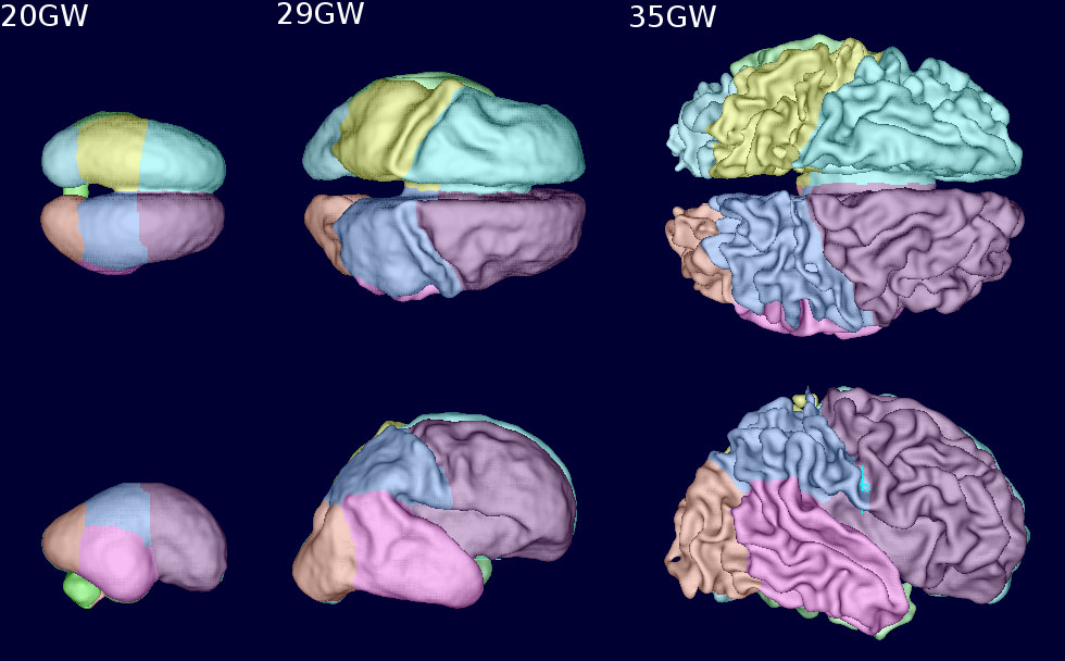
Fetal Brain Growth In The First Trimester Pediagenosis Fetal brain mri is an important contributing tool for fetal vm assessment, providing additive information in 1–14% of cases (primarily related to mcd, callosal, and vermian anomalies) . fetuses with vm should be repeatedly evaluated throughout gestation. Your fetus will begin the process of developing a brain around week 5, but it isn’t until week 6 or 7 when the neural tube closes and the brain separates into three parts, that the real fun begins. The changes that occur in the gross anatomy of the fetal brain reflect dramatic changes occurring at the cellular level. neuron production begins in the embryonic period on e42, and extends through midgestation in most brain areas. regions of the brain that contain the cell bodies of neurons are gray in appearance, hence the name. Early brain development. the human brain is developed through complex processes of neurulation, neuronal prolifertation and migration, apoptosis, and synaptogenesis that start in sequence shorly after conception and progress rapidly before birth. the axons then myelinate and the neocortex matures in a process that completes by adulthood.

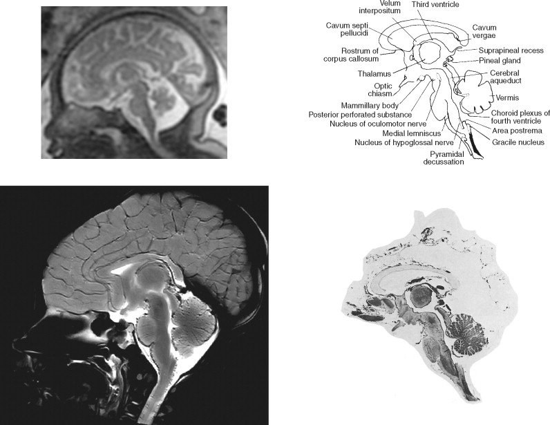
Sectional Anatomy Of The Fetal Brain Radiology Key The changes that occur in the gross anatomy of the fetal brain reflect dramatic changes occurring at the cellular level. neuron production begins in the embryonic period on e42, and extends through midgestation in most brain areas. regions of the brain that contain the cell bodies of neurons are gray in appearance, hence the name. Early brain development. the human brain is developed through complex processes of neurulation, neuronal prolifertation and migration, apoptosis, and synaptogenesis that start in sequence shorly after conception and progress rapidly before birth. the axons then myelinate and the neocortex matures in a process that completes by adulthood. Normal brain development at fetal mri. the knowledge of normal brain development is fundamental for the interpretation of cns fetal anomalies . here follows a synthetic resume of the main processes leading to the formation organization of the fetal brain which can be monitored at fetal mri spatial resolution scale: these processes are disrupted. She is working to link patterns of early brain activity to childhood behavioral outcomes, including speech, motor skills, and cognition. if maps of functional connectivity in the fetal brain turn out to predict health outcomes later in life, the findings will bring us closer to understanding the origins of neurodevelopmental problems.

Comments are closed.