Figure 031 4288 Dual Energy X Ray Absorptiometry Dxa Showing The

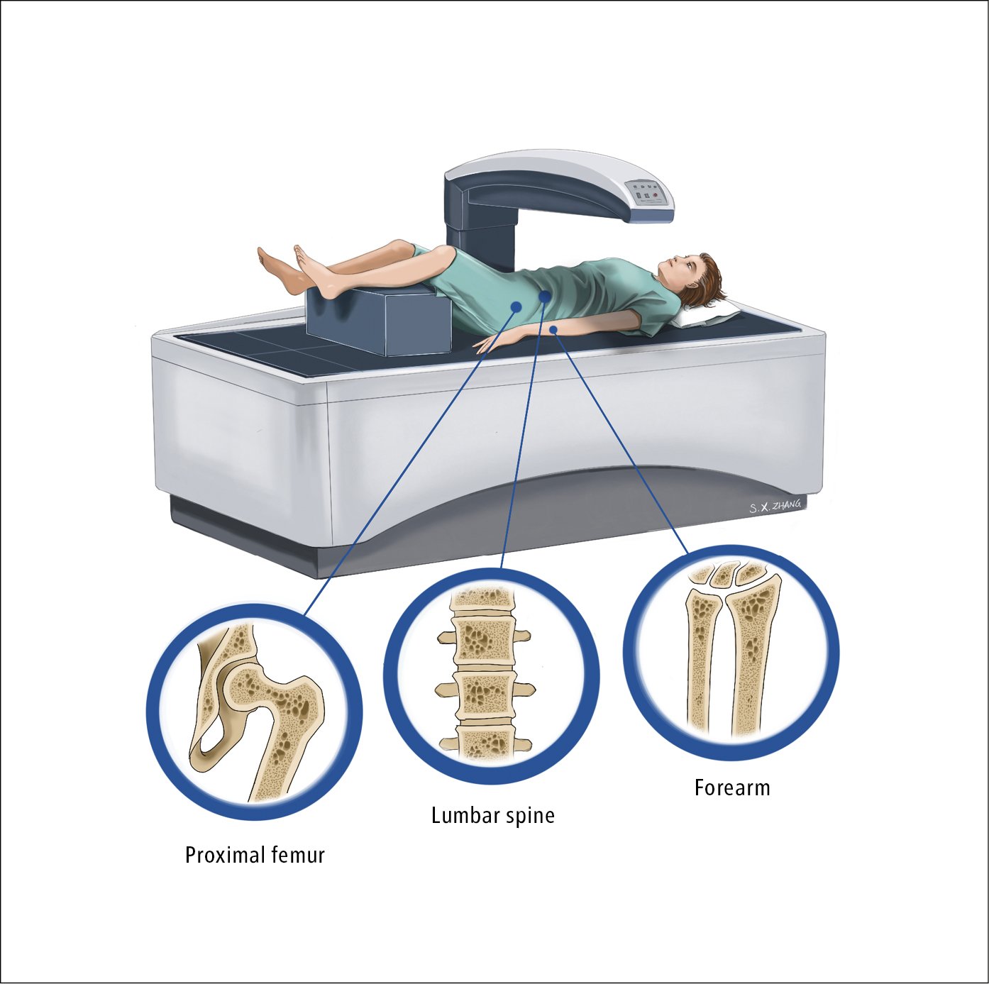
Figure 031 4288 Dual Energy X Ray Absorptiometry Dxa Showing The Figure 031 4288. dual energy x ray absorptiometry (dxa) showing the proximal femur, lumbar spine, and bones of the forearm. illustration courtesy of dr shannon zhang. 20 to 40 mins. dual energy x ray absorptiometry known as : and . bone density scanning and. bone densitometry. what does dexa scan used to measure. bone loss or bone mineral density (bmd) dexa scan determines. bone thickness and strength. risk of fx d t osteoporosis.

Dual Energy X Ray Absorptiometry вђў Bodybuilding Wizard Study with quizlet and memorize flashcards containing terms like dual energy x ray absorptiometry (dxa), a. uses radioactive gadolinium b. is performed on a ct scanner c. uses two discrete gamma photon energies d. is the current standard of care for osteoporosis detection, eighty percent of human skeletal mass is made up of a. cancellous bone b. cortical bone c. spongy bone d. trabecular bone. A dxa scan is an imaging test that measures the strength of your bones. it uses x rays to measure your bone density. dxa is an abbreviation for dual energy x ray absorptiometry. healthcare providers sometimes refer to dxa scans as bone density scans, dxa scans or bone density tests. all of these are different names that refer to the same test. Introduction. in 2017, dual energy x ray absorptiometry (dxa) celebrated its 30 th anniversary ().since the initial development of this technique as the successor of dual photon absorptiometry, dxa technology assisted to continuous technological innovations and is now widely considered as the standard of reference in assessing bone mineral density (bmd) and fracture risk (). Dual energy x ray absorptiometry (dexa) is the technique of choice in the assessment of bone mineral density (bmd) [ 1 ], the average concentration of mineral in a defined section of bone [ 2 ]. dexa is a quick method that is accurate (exact measurement of bmd), precise (reproducible), and flexible (different regions can be scanned) and is.

Dual Energy X Ray Absorptiometry Dxa Positioning And Size Artifacts Introduction. in 2017, dual energy x ray absorptiometry (dxa) celebrated its 30 th anniversary ().since the initial development of this technique as the successor of dual photon absorptiometry, dxa technology assisted to continuous technological innovations and is now widely considered as the standard of reference in assessing bone mineral density (bmd) and fracture risk (). Dual energy x ray absorptiometry (dexa) is the technique of choice in the assessment of bone mineral density (bmd) [ 1 ], the average concentration of mineral in a defined section of bone [ 2 ]. dexa is a quick method that is accurate (exact measurement of bmd), precise (reproducible), and flexible (different regions can be scanned) and is. Whole body dual energy x ray absorptiometry (dxa) is a sensitive and accurate method for quantifying body composition, including both fat mass and lean body mass as a surrogate measure of skeletal. Dual energy x ray absorptiometry (dexa or dxa) is a technique used to aid in the diagnosis of osteopenia and osteoporosis radiographic features. values are calculated for the lumbar vertebrae and femur preferentially, and if one of those sites is not suitable (e.g. artifact, patient mobility), or if there is a history of hyperparathyroidism, the forearm can be used 1.

Dual Energy X Ray Absorptiometry Dxa Iaea Whole body dual energy x ray absorptiometry (dxa) is a sensitive and accurate method for quantifying body composition, including both fat mass and lean body mass as a surrogate measure of skeletal. Dual energy x ray absorptiometry (dexa or dxa) is a technique used to aid in the diagnosis of osteopenia and osteoporosis radiographic features. values are calculated for the lumbar vertebrae and femur preferentially, and if one of those sites is not suitable (e.g. artifact, patient mobility), or if there is a history of hyperparathyroidism, the forearm can be used 1.

Comments are closed.