Illustrations Lymph Node General Histology

Lymph Node Histology Diagram Lymph node enlargement is considered significant based on how large the nodes get. a general rule of thumb states that any enlargement greater than 1 cm of more than one lymph node is considered lymphadenopathy. however, some clinicians say that enlargement should be related to the region the nodes are located in. in other words:. Gross anatomy. lymph nodes are bean shaped structures about 0.1 – 2.5 cm in length. the node is enclosed in a capsule and has an indentation on one surface (along one of its long axes) known as the hilum. the hilum is the point at which arteries carrying nutrients and lymphocytes enter the lymph node and veins leave it.

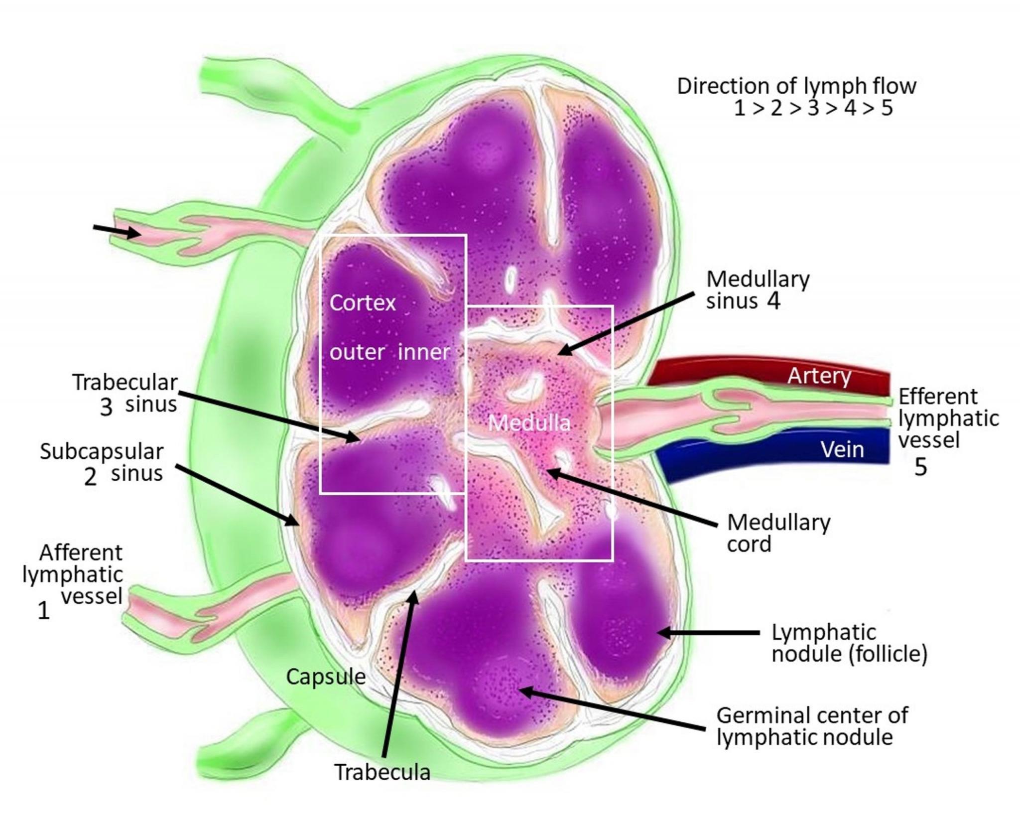
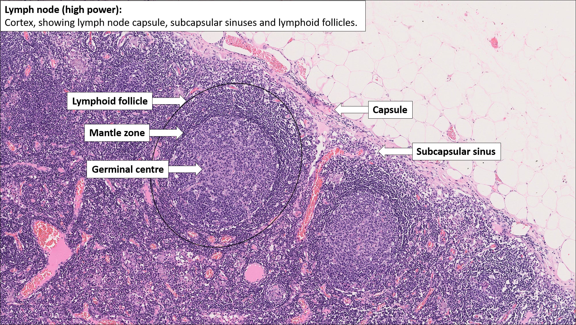
Lymph Node Labeled Histology The lymphoid system is a crucial component of the immune system, encompassing the spleen, thymus, tonsils and lymph nodes. it plays a pivotal role in defending the body against infections by producing and circulating lymphocytes. additionally, the lymphoid system helps maintain fluid balance by returning excess fluid from tissues back into the. Lymph nodes are small organs interposed along lymphatic vessels that immunologically monitor lymph. dense connective tissue enclosing the node. space underneath the capsule that receives lymph from afferent lymphatic vessels. connective tissue that extends inward from the capsule. Scattered throughout the body, lymph nodes perform the critical function of filtering both exogenous and endogenous antigens and producing immune response through activated lymphocytes and antibodies. they can become enlarged in infections, autoimmune diseases, lymphomas, and metastatic carcinomas.[1] histological assessment is imperative in diagnosing these entities. in the case of metastatic. The lymphatic system is composed of lymphatic vessels and lymphoid organs such as the thymus, tonsils, lymph nodes, and spleen. these assist in acquired and innate immunity, in filtering and draining the interstitial fluid, and recycling cells at the end of their life cycle. the fluid that leaks from end stage capillaries returns to the vascular system via the superficial and deep lymphatic.

Histology Lymph Node Lymph Node Histology Slides Lymph Scattered throughout the body, lymph nodes perform the critical function of filtering both exogenous and endogenous antigens and producing immune response through activated lymphocytes and antibodies. they can become enlarged in infections, autoimmune diseases, lymphomas, and metastatic carcinomas.[1] histological assessment is imperative in diagnosing these entities. in the case of metastatic. The lymphatic system is composed of lymphatic vessels and lymphoid organs such as the thymus, tonsils, lymph nodes, and spleen. these assist in acquired and innate immunity, in filtering and draining the interstitial fluid, and recycling cells at the end of their life cycle. the fluid that leaks from end stage capillaries returns to the vascular system via the superficial and deep lymphatic. Text: junquiera, ch 14: the immune system & lymphoid organs. overview: the goal of this lab is to examine the organization of the major organs of the lymphatic system. by the end of the lab, you should be able to describe and distinguish lymph nodules, tonsil, lymph nodes, thymus, and spleen using the criteria given in the table below. Mhs 231 lymph nodes. are small organs interposed along lymphatic vessels. dense connective tissue enclosing the node. space underneath the capsule that receives lymph from afferent lymphatic vessels. outer region of the node. region adjacent to the capsule.

Labeled Lymph Node Histology Diagram Text: junquiera, ch 14: the immune system & lymphoid organs. overview: the goal of this lab is to examine the organization of the major organs of the lymphatic system. by the end of the lab, you should be able to describe and distinguish lymph nodules, tonsil, lymph nodes, thymus, and spleen using the criteria given in the table below. Mhs 231 lymph nodes. are small organs interposed along lymphatic vessels. dense connective tissue enclosing the node. space underneath the capsule that receives lymph from afferent lymphatic vessels. outer region of the node. region adjacent to the capsule.

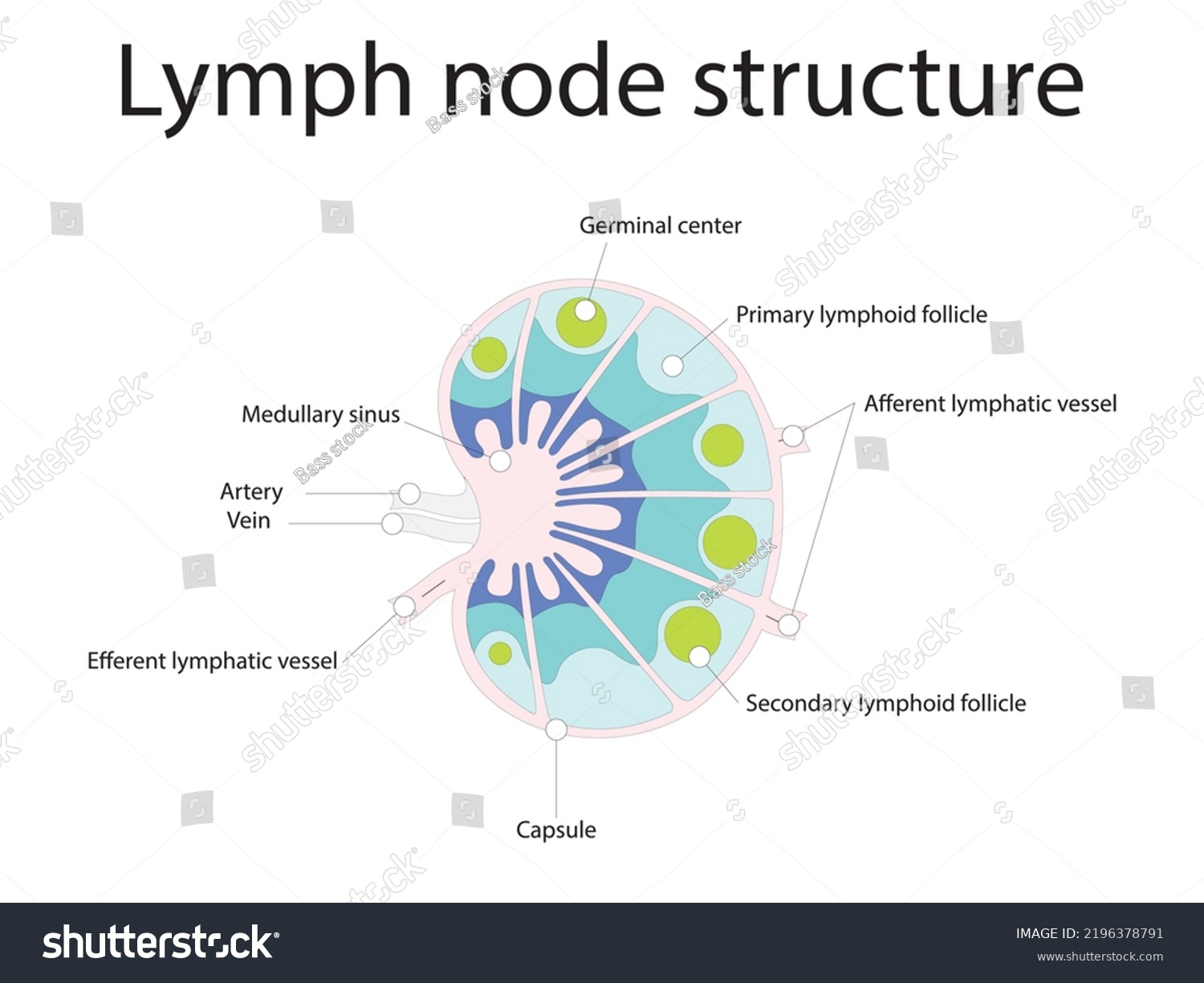
Lymph Node Structure Schematic Anatomic Illustration Stock Vector

Comments are closed.