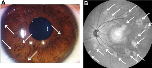
Lisch Nodules Anatomybox

Lisch Nodules Anatomybox Waardenburg, in 1918 first described the pigmented iris hamartomas. karl lisch, an austrian ophthalmologist, reported the association of these iris hamartomas with neurofibromatosis type 1 (nf1) in 1937.[1] the term lisch nodule was used first by riccardi in 1981 in a formal publication. lisch nodules (lns) are the melanocytic hamartomas found in 90% to 100% of adults with nf1 and are the most. Anatomybox.

Lisch Nodule Wikidoc Lisch nodule. other names. iris hamartoma. lisch nodules on surface of iris. lisch nodule, also known as iris hamartoma, is a pigmented hamartomatous nodular aggregate of dendritic melanocytes affecting the iris, [1] named after austrian ophthalmologist karl lisch (1907–1999), who first recognized them in 1937. [2]. They are usually bilateral although unilateral lisch nodules have also been sparsely reported. nf 1 can also impact many different areas of the eye though most are rarer than others. these findings include lisch nodules, optic and or brainstem gliomas, development of glaucoma, astrocytic hamartomas & capillary hemangiomas of the retina, and plexiform neurofibromas. Neurofibromatosis type 1 diagnosis and treatment. Multiple lisch nodules appear to be found only in patients with peripheral neurofibromatosis (neurofibromatosis type 1, or von recklinghausen's disease), an autosomal disorder with a prevalence of.

Morphological Presentations Of Lisch Nodules The Upper And Background Neurofibromatosis type 1 diagnosis and treatment. Multiple lisch nodules appear to be found only in patients with peripheral neurofibromatosis (neurofibromatosis type 1, or von recklinghausen's disease), an autosomal disorder with a prevalence of. Lisch nodules may appear as dome shaped, yellow or brown coloured lesions on iris, best identified on slit lamp examination. 1, 4, 5 they usually precede the appearance of neurofibroma and has a higher prevalence than the latter in younger nf1 patients. 5 these nodules may appear early in childhood and their prevalence and number increases with age. 4, 5 although lisch nodules are reported in. Electronic issn 1533 4406. print issn 0028 4793. the content of this site is intended for health care professionals. a 40 year old man with a history of neurofibromatosis type 1 presented for a.

Comments are closed.