Lisch Nodules Ophthalmology Eyedoc

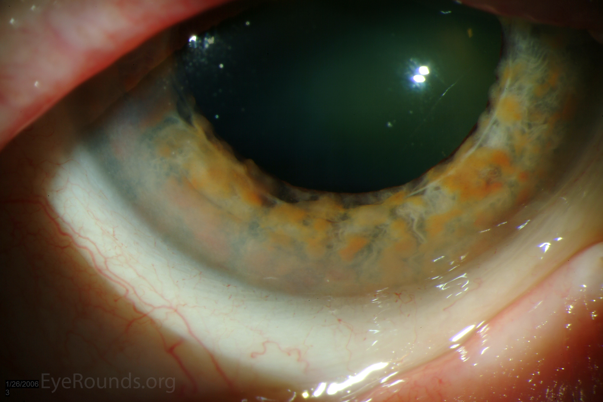
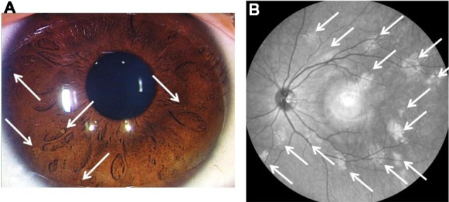
Lisch Nodules Anatomybox Waardenburg, in 1918 first described the pigmented iris hamartomas. karl lisch, an austrian ophthalmologist, reported the association of these iris hamartomas with neurofibromatosis type 1 (nf1) in 1937.[1] the term lisch nodule was used first by riccardi in 1981 in a formal publication. lisch nodules (lns) are the melanocytic hamartomas found in 90% to 100% of adults with nf1 and are the most. A 36 year old woman, with a known case of neurofibromatosis type 1 (nf 1), was observed during her regular ophthalmic examination. anterior segment examination revealed bilateral extensive lisch nodules over the iris (figure). lisch nodules are melanocytic hamartomas of neural crest origin, often associated with nf 1. they are usually bilateral, elevated from the iris surface, tan in.

Atlas Entry Lisch Nodules Lisch nodules – pigmented iris hamartomas; lisch nodules are benign, elevated, tan colored iris nodules considered pathognomonic of nf 1. rarely they are also seen in segmental neurofibromatosis and watson syndrome. they are usually bilateral although unilateral lisch nodules have also been sparsely reported. Lisch nodules are the most common ocular clinical finding in adults older than 20 years with nf 1. unlike café au lait spots, multiple nodules are specific for peripheral nf nf 1 (see the image below). lisch nodules are generally absent in central nf nf 2 and have the following characteristics:. Neurofibromatosis type 1 – lisch nodules (iris pigment epithelium hamartomas) neurofibromatosis type 2 – lisch nodules and skin lesions are less common in nf2 than in nf1. 39. what are the ocular or cns manifestations of neurofibromatosis type 1? 1. neurofibroma 2. lisch nodules (iris pigment epithelium hamartomas) 3. optic nerve glioma 4. The ciliary body may show thickening, and there may be signs of angle infiltration by neurofibroma or lisch nodules. 31 – 33 glaucoma evident at birth usually suggests congenital abnormality of the angle, while onset at a later date suggests involvement of the anterior angle secondary to nf1 15, 35, 36 (figure 4).

Lisch Nodule Wikidoc Neurofibromatosis type 1 – lisch nodules (iris pigment epithelium hamartomas) neurofibromatosis type 2 – lisch nodules and skin lesions are less common in nf2 than in nf1. 39. what are the ocular or cns manifestations of neurofibromatosis type 1? 1. neurofibroma 2. lisch nodules (iris pigment epithelium hamartomas) 3. optic nerve glioma 4. The ciliary body may show thickening, and there may be signs of angle infiltration by neurofibroma or lisch nodules. 31 – 33 glaucoma evident at birth usually suggests congenital abnormality of the angle, while onset at a later date suggests involvement of the anterior angle secondary to nf1 15, 35, 36 (figure 4). Discussion. lisch nodules are the most common ophthalmologic manifestation of nf1, reported in up to 73–95% of cases. 4, 5 they are melanocytic iris hamartomas with no ophthalmologic complications. 1 first reported by waardenburg in 1918, lisch confirmed its association with nf in 1937. 5 unlike other pigmentary abnormalities such as café au lait spots and axillary freckling, multiple lisch. Photographer: ed heffron, brice critser, cra. lisch nodules are melanocytic hamartomas of the iris, often associated with neurofibromatosis (nf) i. they are usually elevated and tan in appearance. their incidence in nf1 increases with age and their prevalence raises by about 10% per year of life, up to age 9. these photographs show the various.

Comments are closed.