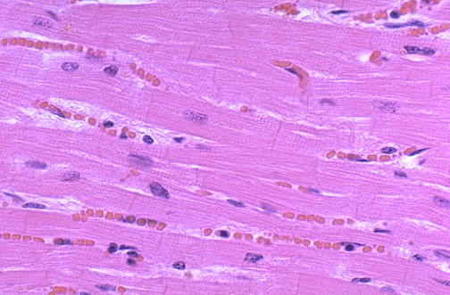
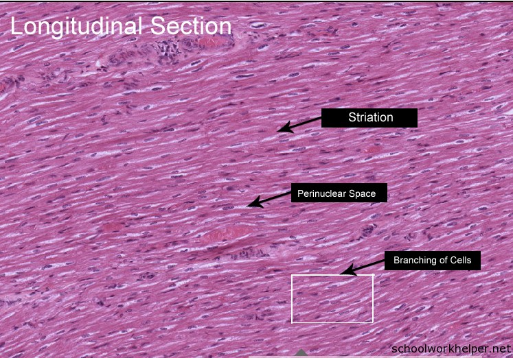
Longitudinal Section Cardiac Muscle Histology Labeled Normal

Normal Histology Characteristics. cardiac muscle tissue, also known as myocardium, is a structurally and functionally unique subtype of muscle tissue located in the heart, that actually has characteristics from both skeletal and muscle tissues. it is capable of strong, continuous, and rhythmic contractions that are automatically generated. Cardiac muscle is striated, involuntary muscle found in the heart wall. cardiac muscle cells (or cardiomyocytes) contain the same contractile filaments as in skeletal muscle. cross section. cardiac muscle cells. have rounded cross sections (less than 25 µm in diameter) with a centrally located nucleus. longitudinal section.
:watermark(/images/watermark_5000_10percent.png,0,0,0):watermark(/images/logo_url.png,-10,-10,0):format(jpeg)/images/overview_image/1902/PoghnLCNI8cl42YWuhwg_histology-cardiac-muscle_english.jpg)
Cardiac Muscle Tissue Histology Kenhub Normal histology. normal cardiac muscle in longitudinal section shows a syncytium of myocardial fibers with central nuclei. faint pink intercalated discs cross some of the fibers. red blood cells are seen in single file in capillaries between the fibers. pathology, medical education, student. The cardiac muscle is a specialized type of involuntary muscle tissue that makes up the bulk of the heart. cardiac muscle cells, or cardiomyocytes, are elongated and spindle shaped, with one to two centrally located nuclei. these cell fibers are arranged in a branching network and intercalated discs, which are junctions that allow these muscle. Cardiac muscle cells possess specialized cell junctions called intercalated discs, and these are visible in longitudinal sections of cardiac muscle. figure 6 5 is a photomicrograph of cardiac muscle in longitudinal section. note that cardiac muscle usually has only one centrally located nucleus per cell. Part i: heart. cardiac muscle fibers can be seen in both cross and longitudinal sections. measure fiber diameters and note blood vessels filled with rbcs between the fibers. fine details of the myocardiocytes can be seen, especially myofibrils, cross striations, intercalated disks, and mitochondria next to the nuclei.

Cardiac Muscle Longitudinal Section Slide Labelled Histology Cardiac muscle cells possess specialized cell junctions called intercalated discs, and these are visible in longitudinal sections of cardiac muscle. figure 6 5 is a photomicrograph of cardiac muscle in longitudinal section. note that cardiac muscle usually has only one centrally located nucleus per cell. Part i: heart. cardiac muscle fibers can be seen in both cross and longitudinal sections. measure fiber diameters and note blood vessels filled with rbcs between the fibers. fine details of the myocardiocytes can be seen, especially myofibrils, cross striations, intercalated disks, and mitochondria next to the nuclei. 43 cardiac muscle view virtual em slide cardiac muscle (longitudinal section). note central location of muscle nuclei. note the "stacks" of mitochondria between myofibrils. cardiac muscle is even richer than skeletal muscle in mitochondria (again, important for energy production). an intercalated disc is present in the upper left region of the. Cardiac muscle (myocardium) is striated, involuntary muscle found in the heart wall. cardiac muscle cells (cardiomyocytes) contain the same contractile filaments as in skeletal muscle (sarcomeres). they are intermediate in size compared to skeletal and smooth muscle. cardiac muscle cells have rounded cross sections with a.

Man Cardiac Muscle Longitudinal Section 500x Man Mammals 43 cardiac muscle view virtual em slide cardiac muscle (longitudinal section). note central location of muscle nuclei. note the "stacks" of mitochondria between myofibrils. cardiac muscle is even richer than skeletal muscle in mitochondria (again, important for energy production). an intercalated disc is present in the upper left region of the. Cardiac muscle (myocardium) is striated, involuntary muscle found in the heart wall. cardiac muscle cells (cardiomyocytes) contain the same contractile filaments as in skeletal muscle (sarcomeres). they are intermediate in size compared to skeletal and smooth muscle. cardiac muscle cells have rounded cross sections with a.

Comments are closed.