Muscles Of Neck Anterior View Anatomy Pediagenosis

Muscles Of Neck Anterior View Anatomy Pediagenosis When the muscles of both sides act together, they flex the neck. innervation: accessory nerve (cn xi and c2 and c3). comment: when the head is fixed, the 2 muscles acting together can help elevate the thorax during forced inspiration. the sternocleidomastoid (scm) is 1 of 2 muscles innervated by the spinal accessory nerve. Muscles of pharynx: medial view anatomy medial pterygoid plate, cartilaginous part of auditory tube (eustachian), tensor veli palatini muscle, pharyngobasilar fascia, levator veli palatini muscle, palatine aponeurosis and tendon of tensor veli palatini muscle, pharyngeal tubercle (basilar part of occipital bone), pharyngeal raphe, anterior longitudinal ligament, anterior atlantooccipital.

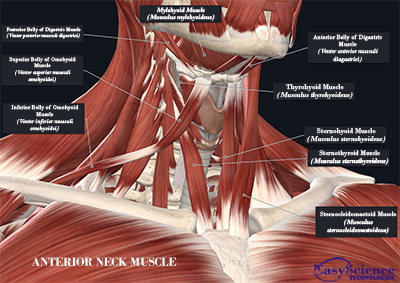
Neck Muscles Anterior Learn Muscles Muscles of the neck (musculi cervicales) the muscles of the neck are muscles that cover the area of the neck. these muscles are mainly responsible for the movement of the head in all directions. they consist of 3 main groups of muscles: anterior, lateral and posterior groups, based on their position in the neck. The muscles of the neck are present in four main groups. the suboccipital muscles act to rotate the head and extend the neck. rectus capitis posterior major and rectus capitis posterior minor attach the inferior nuchal line of the occiput to the c2 and c1 vertebrae respectively. obliquus capitis superior also extends from the occiput to c1. The content of the neck is grouped into 4 neck spaces, called the compartments. vertebral compartment: contains cervical vertebrae and postural muscles. visceral compartment: contains glands (thyroid, parathyroid, and thymus), the larynx, pharynx and trachea. two vascular compartments: contain the common carotid artery, internal jugular vein. The next group of muscles of the anterior neck are the infrahyoid muscles, and as their name suggests, these muscles are located inferior to or below the hyoid bone. this group of muscles are also known as the strap muscles due to their flat ribbon like appearance. they comprise the sternohyoid, sternothyroid, thyrohyoid, and omohyoid muscles.

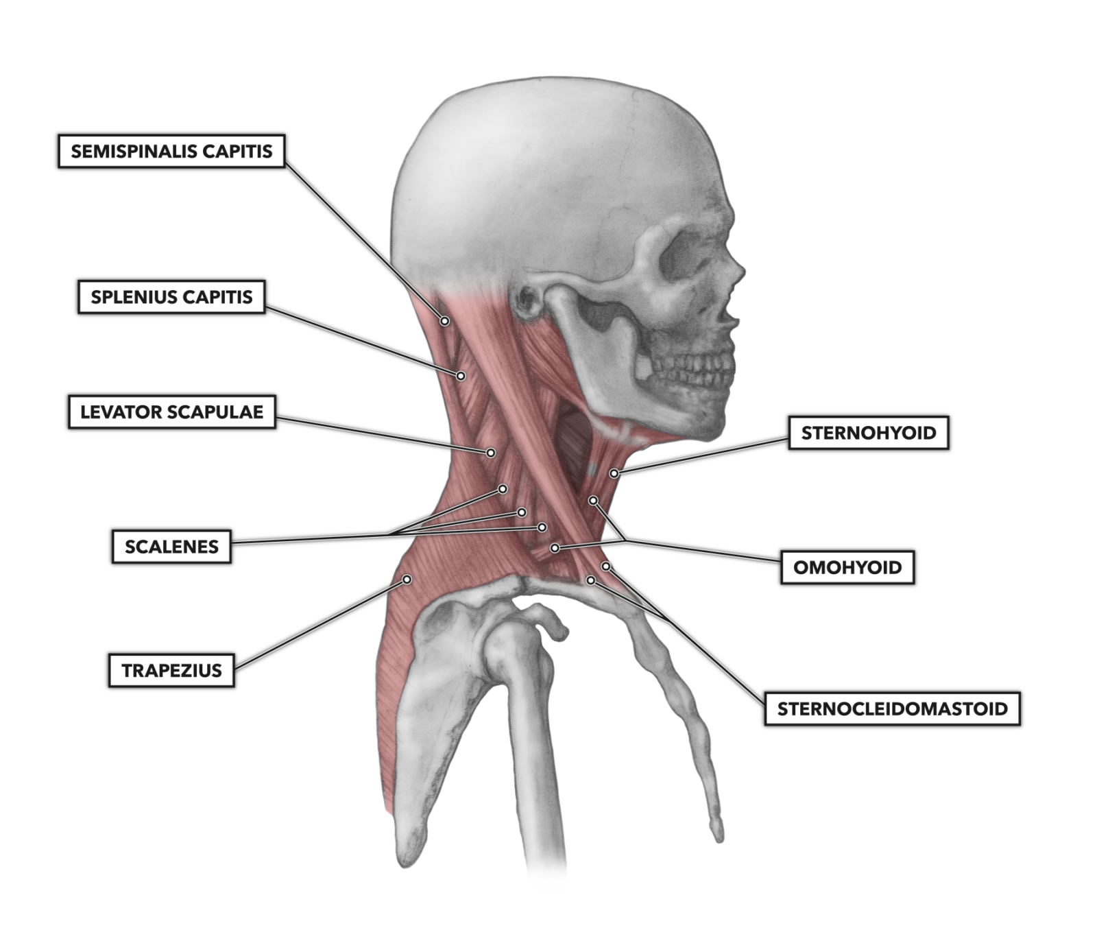
Anterior Neck Muscle вђ Easyscience Technologies The content of the neck is grouped into 4 neck spaces, called the compartments. vertebral compartment: contains cervical vertebrae and postural muscles. visceral compartment: contains glands (thyroid, parathyroid, and thymus), the larynx, pharynx and trachea. two vascular compartments: contain the common carotid artery, internal jugular vein. The next group of muscles of the anterior neck are the infrahyoid muscles, and as their name suggests, these muscles are located inferior to or below the hyoid bone. this group of muscles are also known as the strap muscles due to their flat ribbon like appearance. they comprise the sternohyoid, sternothyroid, thyrohyoid, and omohyoid muscles. The anterior triangle is situated at the front of the neck. it is bounded: superiorly – inferior border of the mandible (jawbone). laterally – anterior border of the sternocleidomastoid. medially – sagittal line down the midline of the neck. investing fascia covers the roof of the triangle, while visceral fascia covers the floor. Scalenes (anterior, middle, and posterior): a group of three muscles at the sides of the neck that side bend and rotate the head. trapezius (traps): a thick neck and shoulder muscle that shrugs the shoulders up and helps the side bend, rotate, and bend the neck backward. levator scapulae: a muscle that travels from the neck on a diagonal down.

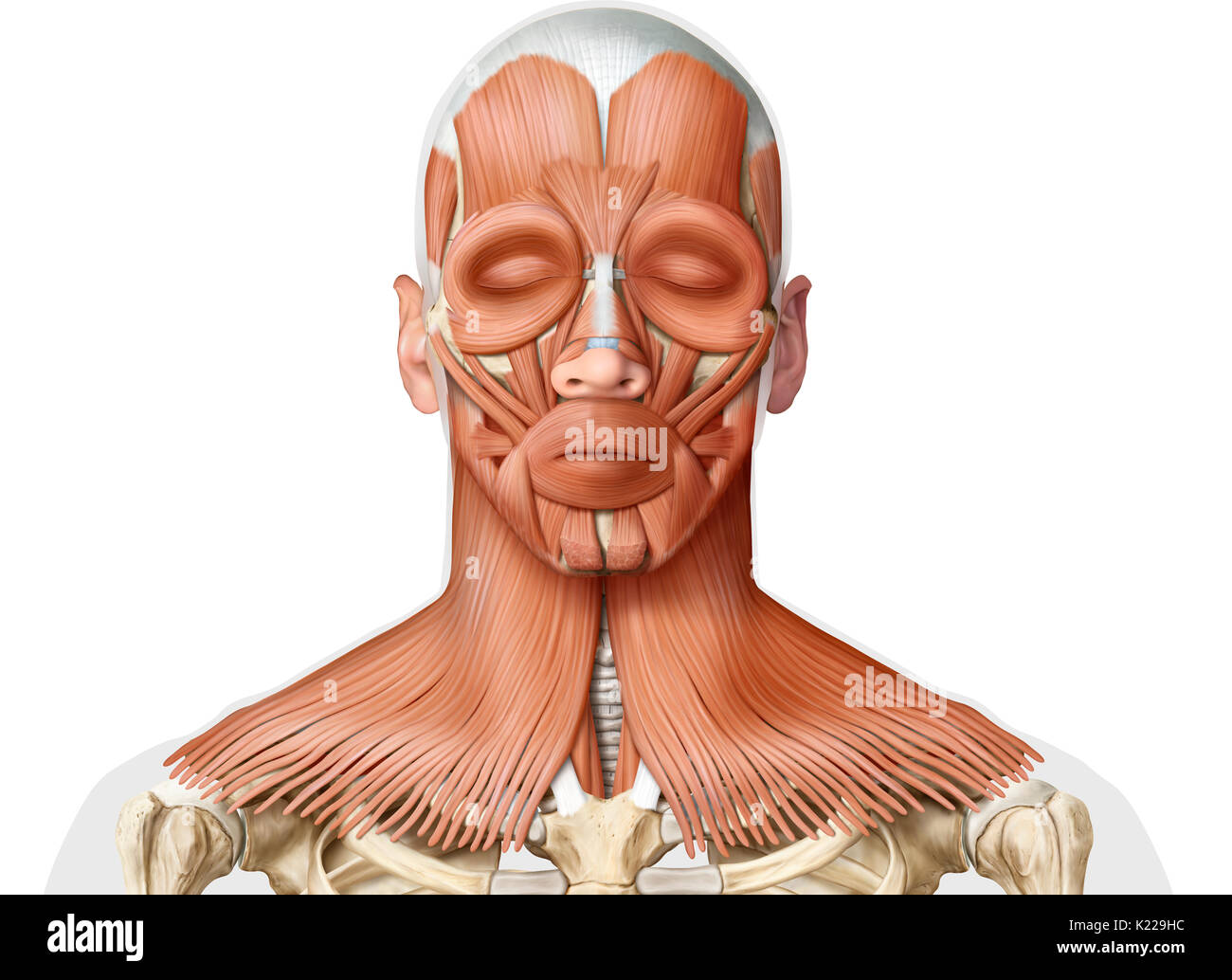
Muscles Anterior Neck Hi Res Stock Photography And Images Alamy The anterior triangle is situated at the front of the neck. it is bounded: superiorly – inferior border of the mandible (jawbone). laterally – anterior border of the sternocleidomastoid. medially – sagittal line down the midline of the neck. investing fascia covers the roof of the triangle, while visceral fascia covers the floor. Scalenes (anterior, middle, and posterior): a group of three muscles at the sides of the neck that side bend and rotate the head. trapezius (traps): a thick neck and shoulder muscle that shrugs the shoulders up and helps the side bend, rotate, and bend the neck backward. levator scapulae: a muscle that travels from the neck on a diagonal down.

Neck Dissection Anatomy Anterior Google Search Painti Vrogue Co

Comments are closed.