Muscles Of Posterior Thoracic Wall Photograph By Askl Vrogue Co

Muscles Of Posterior Thoracic Wall Photograph By Asklepios Medical Science photo library's website uses cookies. muscles of posterior thoracic wall. c020 0418. rights managed. 50.3 mb (50.0 mb compressed) 4193 x 4193 pixels. 1:15 structure of thoracic wall. 2:42 intrinsic muscles of thoracic wall. 6:12 intercostal muscles. 9:25 subcostal muscles. 11:00 extrinsic muscles of thoracic wall. 15:12 clinical notes. 16:23 summary. main muscles found in the thoracic wall. watch the video tutorial now.

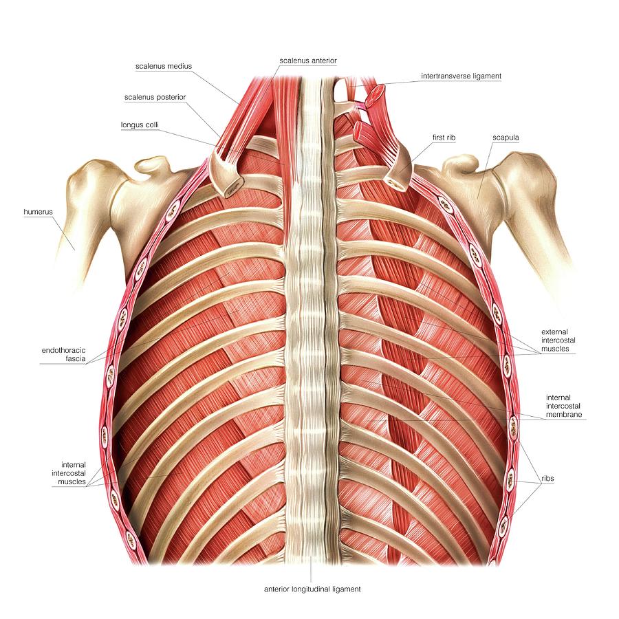
Muscles Of Posterior Thoracic Wall Photograph By Askl Vrogue Co Illustration of posterior thoracic wall. this anterior view labelled illustration is from 'asklepios atlas of the human anatomy'. transform your photos into one of a kind, hand painted masterpieces !. The thoracic, or chest wall, consists of a skeletal framework, fascia, muscles, and neurovasculature – all connected together to form a strong and protective yet flexible cage. the thorax has two major openings: the superior thoracic aperture found superiorly and the inferior thoracic aperture located inferiorly. the superior thoracic. Muscles of the thorax. the muscles of the thorax include both the diaphragm as well as the muscles of the thoracic cage. the diaphragm can be located below the lungs and consists of a sheet of skeletal muscle which displays a double domed structure. the diaphragm is important as it separates the thoracic cavity from the abdominal cavity and. The thoracic (chest) wall is composed of the rib cage, inner and outer muscles, vessels, lymphatics, fascia, and skin. the rib cage is formed by the ribs, costal cartilages, sternum, and thoracic vertebrae. the thoracic inlet is the passage of the trachea, aortic arch arteries, major veins, and lymphatics. the outlet of the thorax is covered by.

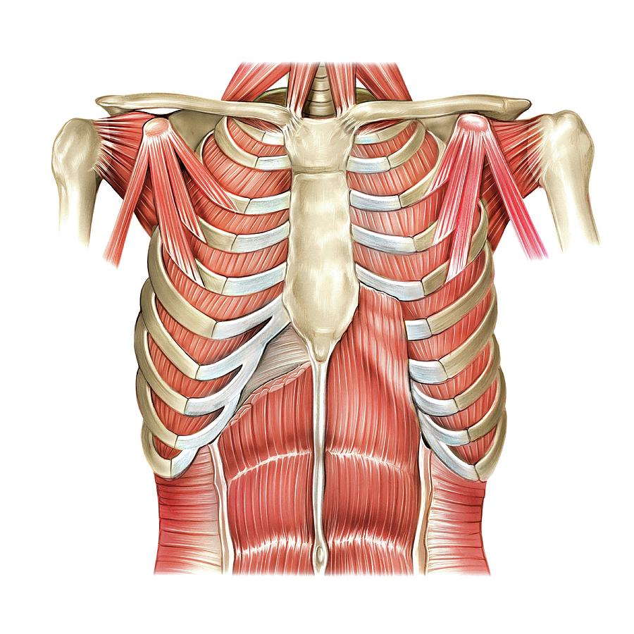
Muscles Of The Thorax Photograph By Asklepios Medical Vrogue Co Muscles of the thorax. the muscles of the thorax include both the diaphragm as well as the muscles of the thoracic cage. the diaphragm can be located below the lungs and consists of a sheet of skeletal muscle which displays a double domed structure. the diaphragm is important as it separates the thoracic cavity from the abdominal cavity and. The thoracic (chest) wall is composed of the rib cage, inner and outer muscles, vessels, lymphatics, fascia, and skin. the rib cage is formed by the ribs, costal cartilages, sternum, and thoracic vertebrae. the thoracic inlet is the passage of the trachea, aortic arch arteries, major veins, and lymphatics. the outlet of the thorax is covered by. Figure 1. a posterior view and b antero lateral view of muscles of the thoracic wall. figure 2. antero lateral view of a subcostal muscles (anterior portion of sternum and ribs dissected out), and b transversus thoracis muscle. figure 3. a tissue cross section of two ribs and associated tissues. There are 11 pairs of external intercostal muscles. they run inferoanteriorly from the rib above to the rib below, and are continuous with the external oblique of the abdomen. attachments: originate at the lower border of the rib, inserting into the superior border of the rib below. actions: elevates the ribs, increasing the thoracic volume.

Muscles Of Posterior Thoracic Wall Photograph By Askl Vrogue Co Figure 1. a posterior view and b antero lateral view of muscles of the thoracic wall. figure 2. antero lateral view of a subcostal muscles (anterior portion of sternum and ribs dissected out), and b transversus thoracis muscle. figure 3. a tissue cross section of two ribs and associated tissues. There are 11 pairs of external intercostal muscles. they run inferoanteriorly from the rib above to the rib below, and are continuous with the external oblique of the abdomen. attachments: originate at the lower border of the rib, inserting into the superior border of the rib below. actions: elevates the ribs, increasing the thoracic volume.

Comments are closed.