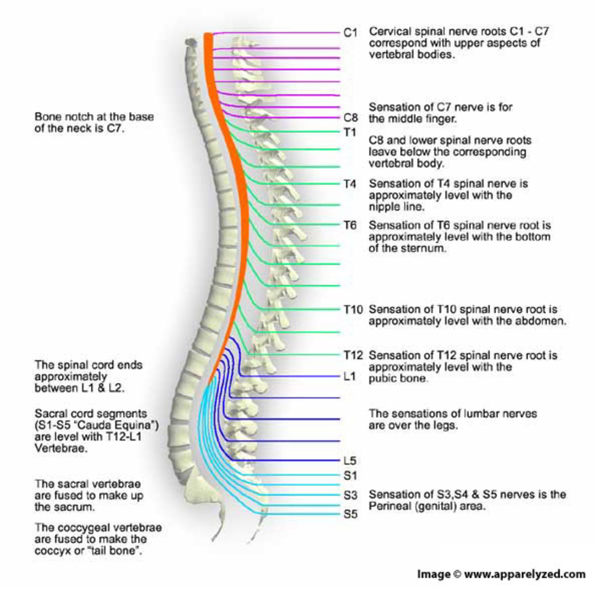
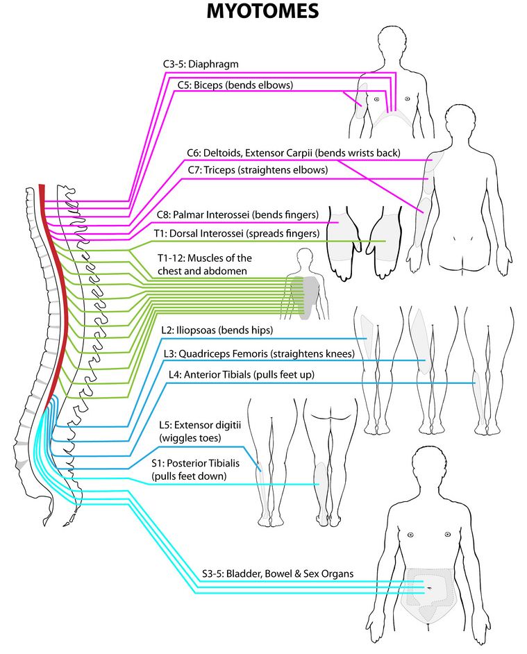
Myotomes Of The Spinal Cord Each Segmental Nerve Root Medical

Myotomes Of The Spinal Cord Each Segmental Nerve Root Grep Myotomes are part of the somatic (voluntary) nervous system, which is part of your peripheral nervous system. the peripheral and central nervous systems communicate with one another. the motor nerve roots are responsible for muscle movement. they branch out from the different parts of the spinal cord. a myotome is the group of muscles on one. A myotome (greek: myo=muscle, tome = a section, volume) is defined as a group of muscles which is innervated by single spinal nerve root. myotome testing is an essential part of neurological examination when suspecting radiculopathy. myotomes are much more complex to test then dermatomes, since each skeletal muscle is innervated by nerves derived from more than one spinal cord level.[1].

Myotomes Of The Spinal Cord Each Segmental Nerve Root Inne Myotomes. anatomy and function of the peripheral nervous system. a myotome is a group of muscles innervated by the ventral root a single spinal nerve. this term is based on the combination of two ancient greek roots; “myo ” meaning “muscle”, and “tome”, a “cutting” or “thin segment”. like spinal nerves, myotomes are. Spinal cord injury (sci) affects conduction of sensory and motor signals across the site (s) of lesion (s), as well as the autonomic nervous system. by systematically examining the dermatomes and myotomes, as described within this booklet, one can determine the cord segments affected by the sci. from the international standards examination. The myotome of a muscle is the basis for diagnosing spinal and peripheral nerve disorders. despite its critical importance in clinical neurology, myotome charts presented in many textbooks, surprisingly, show non negligible discordances with each other. many authors do not even clearly state the bases of their charts. studies that have presented with raw data regarding myotome identification. A dermatome is an area of skin supplied by a single spinal nerve. if you imagine the human body as a map, each dermatome represents the area of skin supplied with sensation by a specific nerve root. it is important to bear in mind that the dermatomes of the head are supplied by branches v1, v2 and v3 of the trigeminal nerve.

The Spinal Cord And Its Importance Owlcation The myotome of a muscle is the basis for diagnosing spinal and peripheral nerve disorders. despite its critical importance in clinical neurology, myotome charts presented in many textbooks, surprisingly, show non negligible discordances with each other. many authors do not even clearly state the bases of their charts. studies that have presented with raw data regarding myotome identification. A dermatome is an area of skin supplied by a single spinal nerve. if you imagine the human body as a map, each dermatome represents the area of skin supplied with sensation by a specific nerve root. it is important to bear in mind that the dermatomes of the head are supplied by branches v1, v2 and v3 of the trigeminal nerve. Shoulder adduction – c678. elbow flexion – c5 (musculocutaneous) elbow extension – c7 (radial) wrist flexion & extension – c67 (radial) finger flexion – c8 (median) finger extension – c7 (radial – posterior interosseous) finger abduction – t1 (ulnar) abductor pollicis brevis – t1 (median) sorting out muscles. Each nerve forms from nerve fibers, known as fila radicularia, extending from the posterior (dorsal) and anterior (ventral) roots of the spinal cord. the roots connect via interneurons. grossly, the root fibers join together within the intervertebral foramina to form a spinal nerve. the dorsal root is composed of afferent sensory axons that.

Spinal Cord Myotomes Shoulder adduction – c678. elbow flexion – c5 (musculocutaneous) elbow extension – c7 (radial) wrist flexion & extension – c67 (radial) finger flexion – c8 (median) finger extension – c7 (radial – posterior interosseous) finger abduction – t1 (ulnar) abductor pollicis brevis – t1 (median) sorting out muscles. Each nerve forms from nerve fibers, known as fila radicularia, extending from the posterior (dorsal) and anterior (ventral) roots of the spinal cord. the roots connect via interneurons. grossly, the root fibers join together within the intervertebral foramina to form a spinal nerve. the dorsal root is composed of afferent sensory axons that.

Comments are closed.