Neck Anatomy Archives Graph Diagram

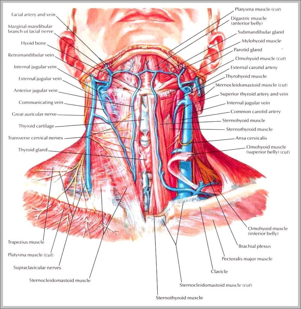
Neck Anatomy Archives Graph Diagram Tag archives: neck anatomy the neck. posted on march 11, 2015 by admin. back of neck anatomy diagram chart diagrams and charts with labels. This human anatomy diagram with labels depicts and explains the details and or parts of the the neck. human anatomy diagrams and charts show internal organs, body systems, cells, conditions, sickness and symptoms information and or tips to ensure one lives in good health.

Head And Neck Anatomy Image Graph Diagram Human neck anatomy teachmeanatomy the neck. The content of the neck is grouped into 4 neck spaces, called the compartments. vertebral compartment: contains cervical vertebrae and postural muscles. visceral compartment: contains glands (thyroid, parathyroid, and thymus), the larynx, pharynx and trachea. two vascular compartments: contain the common carotid artery, internal jugular vein. Neck. the neck is the start of the spinal column and spinal cord. the spinal column contains about two dozen inter connected, oddly shaped, bony segments, called vertebrae. the neck contains seven. Muscles of the neck (musculi cervicales) the muscles of the neck are muscles that cover the area of the neck. these muscles are mainly responsible for the movement of the head in all directions. they consist of 3 main groups of muscles: anterior, lateral and posterior groups, based on their position in the neck.

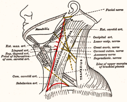
Neck Anatomy Archives Graph Diagram Vrogue Co Neck. the neck is the start of the spinal column and spinal cord. the spinal column contains about two dozen inter connected, oddly shaped, bony segments, called vertebrae. the neck contains seven. Muscles of the neck (musculi cervicales) the muscles of the neck are muscles that cover the area of the neck. these muscles are mainly responsible for the movement of the head in all directions. they consist of 3 main groups of muscles: anterior, lateral and posterior groups, based on their position in the neck. The muscles of the neck are present in four main groups. the suboccipital muscles act to rotate the head and extend the neck. rectus capitis posterior major and rectus capitis posterior minor attach the inferior nuchal line of the occiput to the c2 and c1 vertebrae respectively. obliquus capitis superior also extends from the occiput to c1. Main nerves: maxillary nerve (cn v2), mandibular nerve (cn v3), vagus nerve (cn x), hypoglossal nerve (cn xii), and facial nerve (cn vii) neck. contains hyoid bone, thyroid gland, parathyroid glands, pharynx and larynx; externally divided into triangles, internally divided into compartments. main arteries: common carotid, external carotid.

Comments are closed.