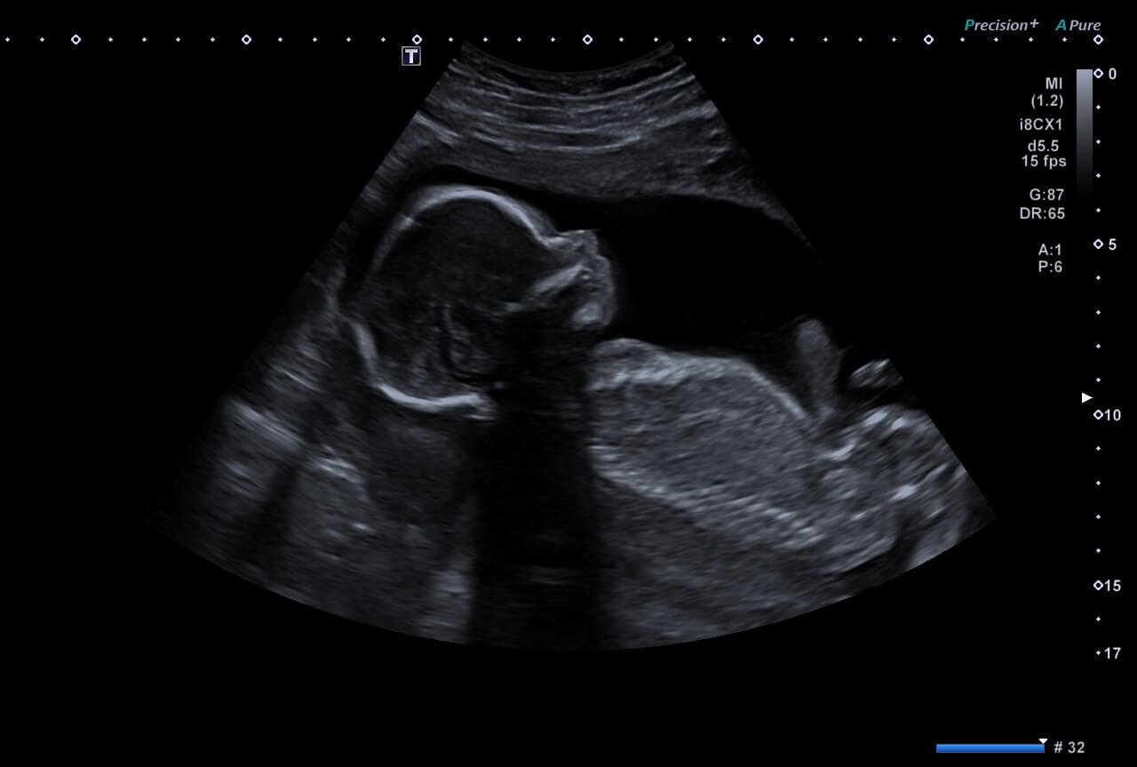
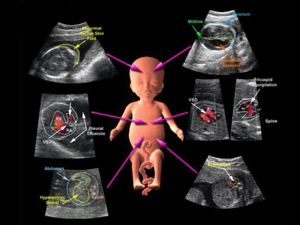
Pin By Julie Boyce On Radiology Pictures Obstetric Ultrasound

Pin By Julie Boyce On Radiology Pictures Obstetric Ultrasound Jan 30, 2021 this pin was discovered by julie boyce. discover (and save!) your own pins on pinterest. jan 30, 2021 this pin was discovered by julie boyce. Jan 12, 2024 this pin was discovered by julie boyce. discover (and save!) your own pins on pinterest.

Pin By Julie Boyce On Abdominal Ultrasound In 2020 Ultrasound Liked by julie boyce. compassionate healthcare professional with clinical experience in general and vascular…. · experience: simonmed imaging · education: west coast ultrasound institute. December 20, 2019march 6, 2020 sonographictendencies obstetrics, ultrasound ob gyn, ob ultrasound, obstetric, sonography, ultrasound. ob ultrasound second trimester (the basics) the second trimester of pregnancy is from week 13 to week 28 – roughly months four, five and six. a second trimester sonogram is usually performed between weeks 18 20. Us remains the imaging method of choice for dating the pregnancy, monitoring fetal growth, assessing fetal well being, and evaluating fetal anatomy and maternal pelvic organs. transvaginal us is particularly useful in the assessment of first trimester pregnancy and in the demonstration of fetal anatomic structures deep in the pelvis. modern us. This pin was discovered by julie boyce. discover (and save!) your own pins on pinterest.

Obstetric Pregnancy Ultrasound Coastal Radiology Us remains the imaging method of choice for dating the pregnancy, monitoring fetal growth, assessing fetal well being, and evaluating fetal anatomy and maternal pelvic organs. transvaginal us is particularly useful in the assessment of first trimester pregnancy and in the demonstration of fetal anatomic structures deep in the pelvis. modern us. This pin was discovered by julie boyce. discover (and save!) your own pins on pinterest. Step 1 – optimize depth to see gestational sac. step 2 – obtain a sagittal view of the gestational sac. step 3 – measure the height and length of the sac using the mean sac diameter calculation package. step 4 – rotate the probe 90º to obtain a transverse view of the gestational sac. The technologic advances in ultrasound imaging, including 3d 4d and volumetric measurements, the use of high frequency transvaginal probes, and the utility for chromosomal screening in early pregnancy (e.g., nuchal translucency) have only expanded the indications for sonographic imaging in the obstetric patient.

Obstetric Ob Ultrasound Made Easy Step By Step Guide Pocus 45 Off Step 1 – optimize depth to see gestational sac. step 2 – obtain a sagittal view of the gestational sac. step 3 – measure the height and length of the sac using the mean sac diameter calculation package. step 4 – rotate the probe 90º to obtain a transverse view of the gestational sac. The technologic advances in ultrasound imaging, including 3d 4d and volumetric measurements, the use of high frequency transvaginal probes, and the utility for chromosomal screening in early pregnancy (e.g., nuchal translucency) have only expanded the indications for sonographic imaging in the obstetric patient.

Obstetric Ultrasound

Comments are closed.