Proximal Superfi Cial Femoral Artery Stenosis Color Fl Ow Ultrasound

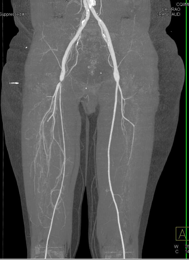
Proximal Superfi Cial Femoral Artery Stenosis Color Fl Ow Ultrasound Duplex image of a severe superficial femoral artery stenosis. the color flow image shows a localized, high velocity jet with color aliasing. spectral waveforms obtained from the site of stenosis indicate peak velocities of more than 400 cm s. sfa prox , proximal superficial femoral artery. Femoral segments were most commonly affected with 43.33% in superficial femoral artery followed by 30% in popliteal artery and 20% in the common femoral artery. below knee involvement was seen in.

High Grade Stenosis Right Femoral Artery Vascular Case Studies Noninvasive spectral doppler waveform assessment is a principal diagnostic tool used in the diagnosis of arterial and venous disease states. with 200 million people affected by peripheral artery disease worldwide 1,2 and >600 000 hospital admissions yearly for venous thromboembolic disease in the united states, 3,4 establishment and adoption of nomenclature for spectral doppler waveform. The steps of color doppler ultrasonography (us) for the lower extremities above the knee, with the patient’s position indicated. the red rectangular boxes are the essential scanning sites and planes for the femoral arteries and the popliteal artery. the numbers within the boxes represent the general steps of scanning. Figure 4. a patient with a normal color flow duplex ultrasound and multiphasic waveforms at the level of the superficial femoral artery (sfa). however, waveforms at the popliteal artery show spectral broadening and the blood velocity (vel) has increased to 576 cm s which indicates that stenosis is present in the popliteal artery. This retrospective study determined the duplex ultrasound scanning criteria for detecting 50%–69% and 70%–99% stenosis of the superficial femoral artery (sfa). examinations of 278 limbs in 185 patients with peripheral arterial disease were performed. duplex ultrasound scanning was used to measure the diameter of the vascular lumen, the peak systolic velocity (psv) at the stenotic segment.

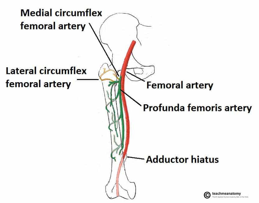
The Hip Joint Articulations Movements Teachmeanatomy Figure 4. a patient with a normal color flow duplex ultrasound and multiphasic waveforms at the level of the superficial femoral artery (sfa). however, waveforms at the popliteal artery show spectral broadening and the blood velocity (vel) has increased to 576 cm s which indicates that stenosis is present in the popliteal artery. This retrospective study determined the duplex ultrasound scanning criteria for detecting 50%–69% and 70%–99% stenosis of the superficial femoral artery (sfa). examinations of 278 limbs in 185 patients with peripheral arterial disease were performed. duplex ultrasound scanning was used to measure the diameter of the vascular lumen, the peak systolic velocity (psv) at the stenotic segment. In this article we focus on lower extremity pad and specifically on the superficial femoral and proximal popliteal artery (sfpa), which are the most common anatomic locations of lower extremity atherosclerosis. we summarize current evidence on the diagnostic evaluation as well as the medical, endovascular and surgical management of sfpa disease. In stent restenosis is a pervasive challenge to the durability of stenting for the treatment of lower extremity ischemia. there is considerable controversy about the criteria for diagnosis, indications for treatment, and preferred algorithm for addressing in stent restenosis. this evidence summary seeks to review existing information on strategies for the treatment of this difficult problem.

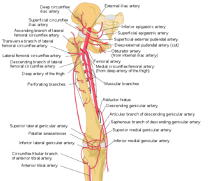
Femoral Artery Physiopedia In this article we focus on lower extremity pad and specifically on the superficial femoral and proximal popliteal artery (sfpa), which are the most common anatomic locations of lower extremity atherosclerosis. we summarize current evidence on the diagnostic evaluation as well as the medical, endovascular and surgical management of sfpa disease. In stent restenosis is a pervasive challenge to the durability of stenting for the treatment of lower extremity ischemia. there is considerable controversy about the criteria for diagnosis, indications for treatment, and preferred algorithm for addressing in stent restenosis. this evidence summary seeks to review existing information on strategies for the treatment of this difficult problem.

Comments are closed.