Rectus Femoris Muscle Injury With Dr Zach Bailowitz Amssm Sports Ultrasound Case Presentation

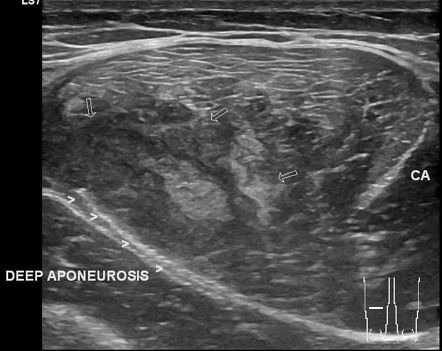
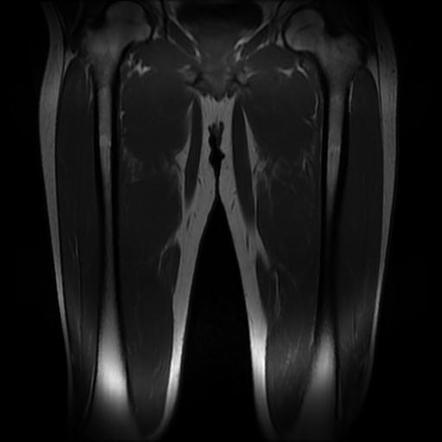
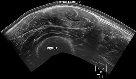
Rectus Femoris Muscle Injury With Dr Zach Bailowitz Ams Dr. zach bailowitz gives an amssm sports ultrasound case presentation about a rectus femoris muscle injury. A rectus femoris strain is a traumatic injury caused by overstretching of the muscle which results in tearing of the muscle fibers of the rectus femoris. diagnosis is made clinically with tenderness over the rectus femoris made worse with resisted hip flexion or extension. treatment is conservative with nsaids, rest and stretching. epidemiology.

Rectus Femoris Muscle Injury Radiology Case Radiopaedia Org The rectus femoris muscle crosses two joints, plays an active part in knee extension and hip flexion and features a high proportion of fast twitch (type ii) muscle fibres and is characterised by a complex musculotendinous architecture, which is considered a predisposing factor for strain injury 1. muscle injury patterns include strains. Rectus femoris (rf) injury is a concern in sports. the management rf strains tears and avulsion injuries need to be clearly outlined. a systematic review of literature on current management strategies for rf injuries, and to ascertain the efficacy thereof by the return to sport (rts) time and re injury rates. Rectus femoris tendon strain symptoms. symptoms include: sudden sharp pain at the front of the hip or groin. swelling and bruising may develop over the site of injury. it will feel particularly tender when pressing in (palpating) where the tendon attaches at the front of the hip. if a complete rupture has occurred then it will be impossible to. The rectus femoris is the anterior thigh compartment's most superficial and nearly vertically oriented muscle. this bipennate structure is a component of the quadriceps muscle complex, one of the knee's most important dynamic stabilizers.[1] the rectus femoris is also known as the "kicking muscle" for its involvement in activities involving forceful knee extension. as in other musculoskeletal.

Rectus Femoris Muscle Injury Radiology Case Radiopaedia Org Rectus femoris tendon strain symptoms. symptoms include: sudden sharp pain at the front of the hip or groin. swelling and bruising may develop over the site of injury. it will feel particularly tender when pressing in (palpating) where the tendon attaches at the front of the hip. if a complete rupture has occurred then it will be impossible to. The rectus femoris is the anterior thigh compartment's most superficial and nearly vertically oriented muscle. this bipennate structure is a component of the quadriceps muscle complex, one of the knee's most important dynamic stabilizers.[1] the rectus femoris is also known as the "kicking muscle" for its involvement in activities involving forceful knee extension. as in other musculoskeletal. The quadriceps muscles are composed of the rectus femoris, vastus lateralis, vastus medialis, and vastus intermedius (picture 1 and figure 1 and figure 2 and figure 3 and figure 4). the rectus femoris lies centrally at the anterior thigh and has two origins . more superficial fibers begin as a tendon at the anterior inferior iliac spine (aiis. —25 year old man with grade iii tear of rectus femoris muscle. longitudinal sonogram (c), obtained in same location as a, and diagram (d) show rectus femoris muscle (rf) in relation to its tendon (t), confirming that tear is at musculotendinous junction. rectus femoris muscle lies superficial to vastus intermedius muscle (vi).

Rectus Femoris Muscle Injury Radiology Reference Article The quadriceps muscles are composed of the rectus femoris, vastus lateralis, vastus medialis, and vastus intermedius (picture 1 and figure 1 and figure 2 and figure 3 and figure 4). the rectus femoris lies centrally at the anterior thigh and has two origins . more superficial fibers begin as a tendon at the anterior inferior iliac spine (aiis. —25 year old man with grade iii tear of rectus femoris muscle. longitudinal sonogram (c), obtained in same location as a, and diagram (d) show rectus femoris muscle (rf) in relation to its tendon (t), confirming that tear is at musculotendinous junction. rectus femoris muscle lies superficial to vastus intermedius muscle (vi).

Comments are closed.