Sem Images Backscattered Electrons And Localized Eds Analyses Wt

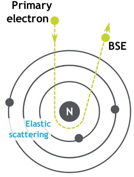
Backscattered Electron Bse Sem Imaging Advancing Materials Sem images (backscattered electrons) and localized eds analyses (wt.%) of polished cross sections through corroded samples (bare zm21, with general view and high magnification detail (a) and peo. Detection of backscattered electrons. the detection of bses is often carried out by detectors that consist of a semiconductor material, typically silicon, placed directly above the sample. electrons that hit the detectors excite the silicon electrons, creating an electron hole pair. semiconductor detectors are only sensitive to electrons with.
What Does Bse Stand For Backscattered Electron вђ Materials Science Ule describes the properties of backscattered electrons and how those properties are modified by specimen characteris tics to produce useful information in sem images. 2.1.1 the numerical measure of backscattered electrons backscattered electrons are quantified with the “backscat tered electron coefficient,” η, defined as η=nn bseb (2.1. For example, backscattered electrons produce images with contrast that carry information about the differences in the atomic number, while secondary electrons produce topographic information about the sample. yet when sem is joined with an edx detector, x rays can also be used as a signal to produce chemical information. to understand how these. In sem, imaging is carried out by using the emission of secondary electrons (topography) and backscattered electrons (atomic number). analytical x rays enable qualitative and quantitative analysis of the specimens. thus, sem provides information on the topography, morphology, composition and crystallographic nature of the analysed specimens. Back scattered electrons (bse) gray levels images were calibrated prior to the run of analyses, in order to obtain consistent brightness and contrast for all the analyzed areas. acquisition with oxford instruments aztec feature ® mode offers the possibility to analyze a frame after the identification of the particles (i.e., the mineral phases.

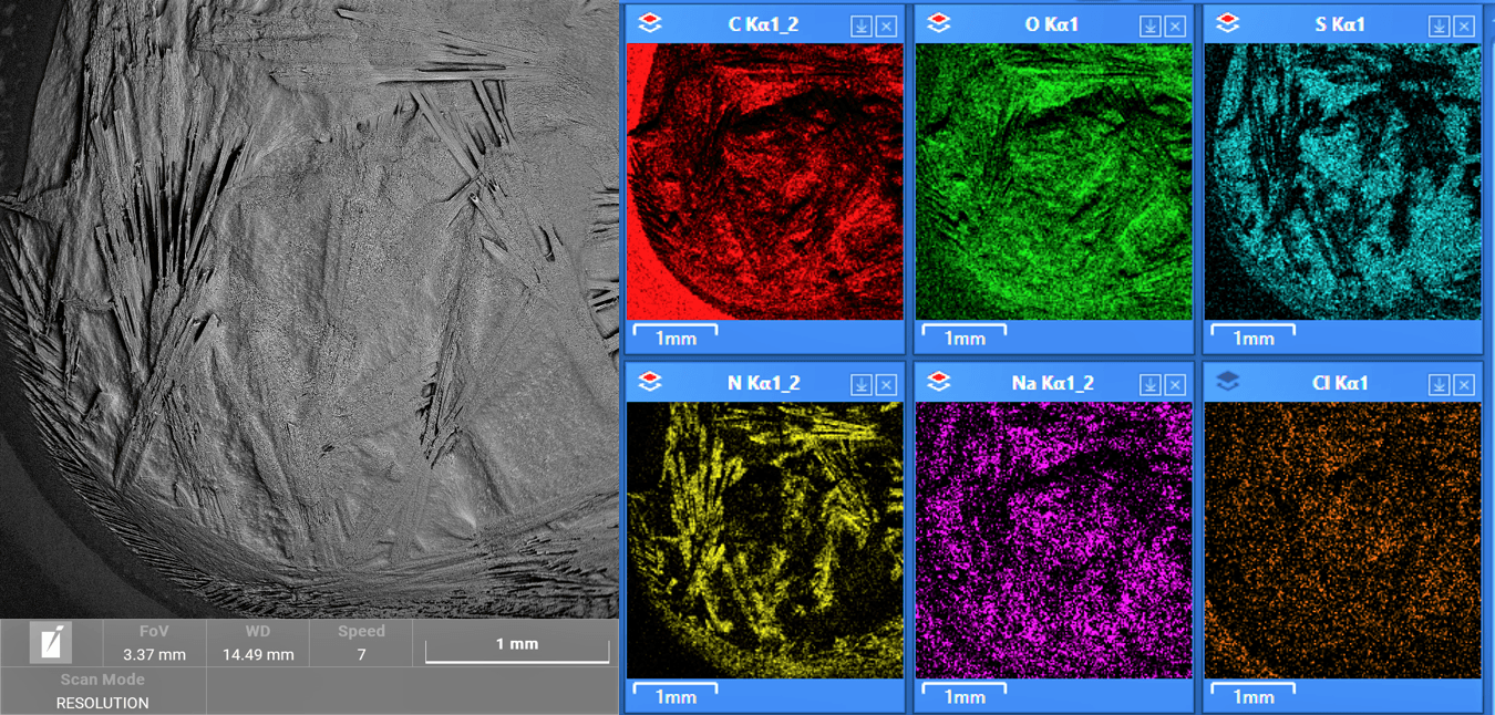
New Sem Eds In sem, imaging is carried out by using the emission of secondary electrons (topography) and backscattered electrons (atomic number). analytical x rays enable qualitative and quantitative analysis of the specimens. thus, sem provides information on the topography, morphology, composition and crystallographic nature of the analysed specimens. Back scattered electrons (bse) gray levels images were calibrated prior to the run of analyses, in order to obtain consistent brightness and contrast for all the analyzed areas. acquisition with oxford instruments aztec feature ® mode offers the possibility to analyze a frame after the identification of the particles (i.e., the mineral phases. 1. introduction. the development of high sensitivity backscattered electron (bse) detectors in scanning electron microscopy (sem) has greatly facilitated the examination of material microstructures across various research domains, such as cementitious science [1], [2], mineralogy identification [3], pavement material [4], and metal structure [5]. Energy dispersive spectroscopy (eds) (supplementary figs. 11 and 12) and additional sem images (supplementary figs. 13–50) have been compiled in the si. results from eds analyses are summarized.

Sem Images Backscattered Electrons And Localized Eds Analyses Wt 1. introduction. the development of high sensitivity backscattered electron (bse) detectors in scanning electron microscopy (sem) has greatly facilitated the examination of material microstructures across various research domains, such as cementitious science [1], [2], mineralogy identification [3], pavement material [4], and metal structure [5]. Energy dispersive spectroscopy (eds) (supplementary figs. 11 and 12) and additional sem images (supplementary figs. 13–50) have been compiled in the si. results from eds analyses are summarized.

Energy Dispersive Spectroscopy On The Scanning Electron Microscope

Comments are closed.