Skin Fungus Under Microscope

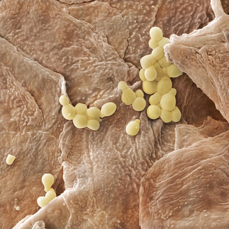
Microscopic Close Up Of A Fungal Infection Of The Upper Skin Layer Dermatopathology and the diagnosis of fungal infections. Skin scrapings and nail clippings are taken to establish or confirm the diagnosis of a fungal infection by microscopy and culture (mycology). scrapings of scale are best taken from the leading edge of the rash after the skin has been cleaned with alcohol. gently remove the surface skin using a blade or curette and place in a sterile container.

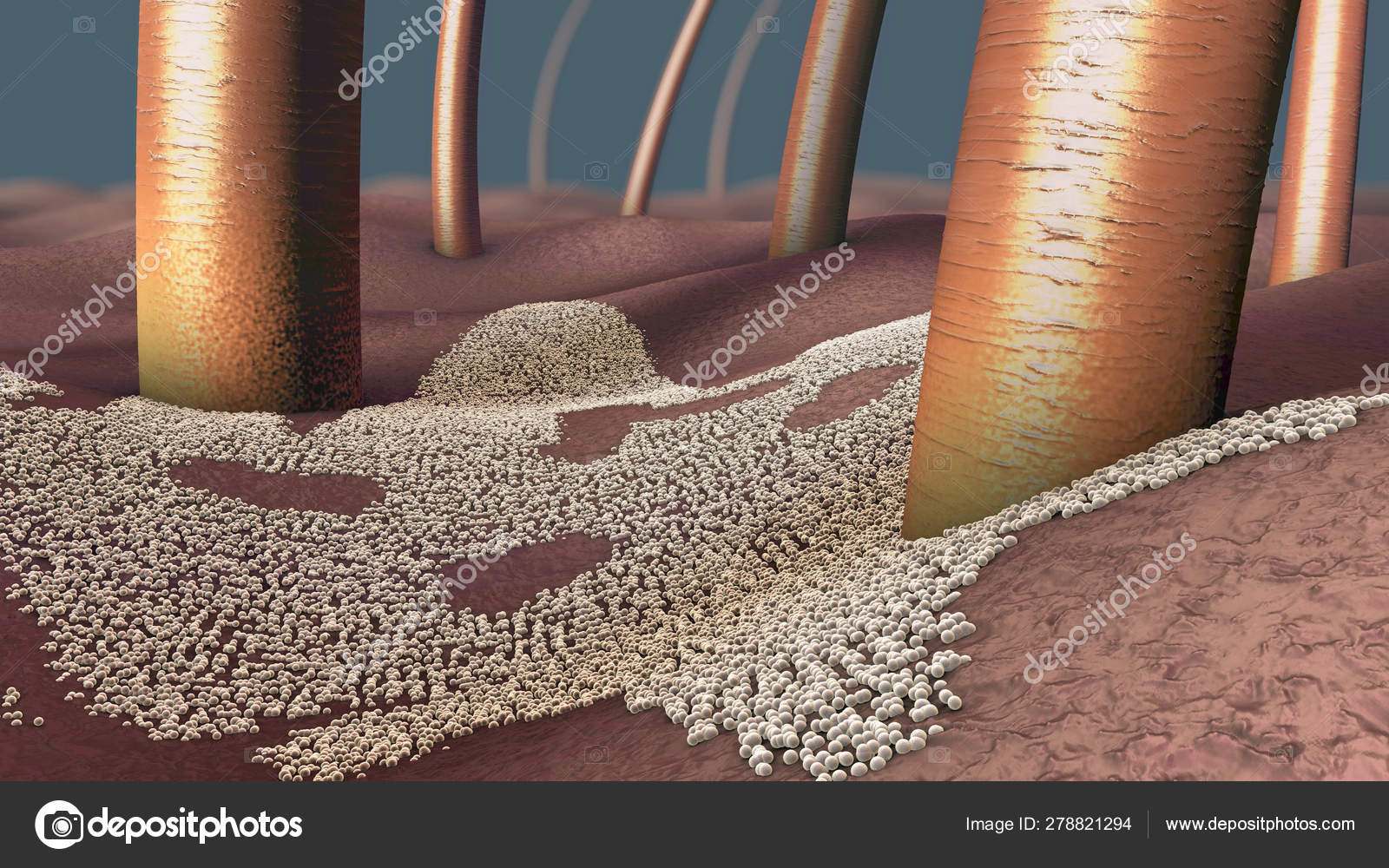
Skin Fungus Under Microscope Histopathologic diagnosis of fungal infections in the 21st. Laboratory tests for fungal infections. Next, the sample is looked at under the microscope, making it is very easy to see if there is a fungus in the sample. how to prepare people do not usually need to prepare in advance for a skin. In most cases, diagnosis can be confirmed by additional diagnostic tests, including direct microscopy, fungal cultures, or wood’s light examination. direct microscopy is typically performed with a potassium hydroxide (koh) preparation that allows the branching filaments of the fungi (hyphae) to be seen under the microscope.

Observation Of Fungi Bacteria And Parasites In Clinical Skin Samples Next, the sample is looked at under the microscope, making it is very easy to see if there is a fungus in the sample. how to prepare people do not usually need to prepare in advance for a skin. In most cases, diagnosis can be confirmed by additional diagnostic tests, including direct microscopy, fungal cultures, or wood’s light examination. direct microscopy is typically performed with a potassium hydroxide (koh) preparation that allows the branching filaments of the fungi (hyphae) to be seen under the microscope. Through a koh examination, superficial fungal infections are easily diagnosed under the microscope by their long branch like structures known as hyphae. to perform a koh examination of the skin and nails, scales or subungual debris are collected by scraping the involved area with a no. 15 blade. Overview of fungal skin infections.

Skin Fungus Under Microscope Through a koh examination, superficial fungal infections are easily diagnosed under the microscope by their long branch like structures known as hyphae. to perform a koh examination of the skin and nails, scales or subungual debris are collected by scraping the involved area with a no. 15 blade. Overview of fungal skin infections.

Microscopic Close Up Of A Fungal Infection Of The Upper Skin Layer

Comments are closed.