Smith Fracture And Scaphoid Tubercle Fracture Radiology Case

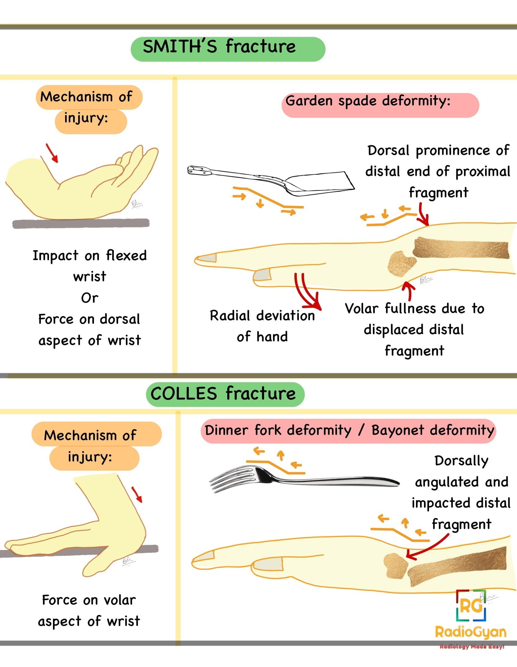
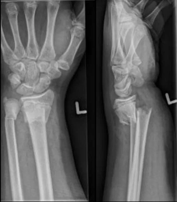
Pin On A G Mcq Age: 25 years. gender: male. x ray. intra articular distal radial fracture with volar displacement (smith fracture). undisplaced scaphoid tubercle fracture. Citation, doi, disclosures and article data. smith fractures, also known as goyrand fractures in the french literature 3, are fractures of the distal radius with associated volar angulation of the distal fracture fragment (s). classically, these fractures are extra articular transverse fractures and can be thought of as a reverse colles fracture.

Smith Fracture Distal Radial Fracture Radiology Case Radiogyan Internal fixation is recommended for all scaphoid waist fractures with dislocation ≥ 1.5 mm. distal scaphoid fractures can be treated conservatively. the majority heal uneventfully after four to six weeks of immobilization, depending on fracture type. in general, proximal scaphoid fractures should be treated with internal fixation. The differential diagnosis for suspected scaphoid injuries includes fractures of other metacarpal bones or the distal radius, scapholunate dissociation, arthritis, tenosynovitis, or strains. The scaphoid is the most commonly fractured bone in the wrist but 20% to 40% of scaphoid fractures are radiographically occult. delayed or misdiagnosis can have significant consequences with late complications such as nonunion, malunion, or the development of avascular necrosis in the proximal pole. after initial negative radiographs, advanced cross sectional imaging, including ct and mri. Anatomy and biomechanics. the scaphoid is a biomechanically important, boat shaped carpal bone (from the greek “skaphos,” meaning “boat”) that articulates with the distal radius, trapezium.

Colle Fracture Distal Radial Fracture Radiology Case Radiogyan The scaphoid is the most commonly fractured bone in the wrist but 20% to 40% of scaphoid fractures are radiographically occult. delayed or misdiagnosis can have significant consequences with late complications such as nonunion, malunion, or the development of avascular necrosis in the proximal pole. after initial negative radiographs, advanced cross sectional imaging, including ct and mri. Anatomy and biomechanics. the scaphoid is a biomechanically important, boat shaped carpal bone (from the greek “skaphos,” meaning “boat”) that articulates with the distal radius, trapezium. Imaging of the scaphoid. there are several different diagnostic modalities to detect a scaphoid fracture. these include conventional radiographs, computed tomography (ct scans), magnetic resonance examination, bone scintigraphy and sonograms. each procedure has its specific advantages and disadvantages (table 1 ). The scaphoid bone is one of 8 carpal bones in the wrist. the scaphoid begins ossification around the 4th year of age and may be earlier in females than males. scaphoid fractures are much more common in adolescents than younger children. approximately 75% of the arterial supply is from branches of the radial artery through vascular perforations.

Comments are closed.