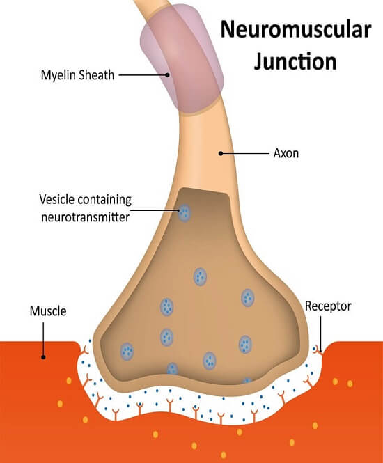
Structure Of A Neuromuscular Junction And Skeletal Muscle Fiber

Anatomy Of Neuromuscular Junction Medicinebtg At its simplest, the neuromuscular junction is a type of synapse where neuronal signals from the brain or spinal cord interact with skeletal muscle fibers, causing them to contract. the activation of many muscle fibers together causes muscles to contract, which in turn can produce movement. the neuromuscular junction then, is a key component in. The “structural and functional” aspects of muscle, starting with the three most basic units that drive skeletal muscle contraction, namely (a) neuromuscular junction (nmj) which serves as a junction between nerve and muscle; (b) machinery involved in excitation–contraction coupling (ecc), which is the process of transduction of electric impulses from nerve to muscle, required to initiate.

Structure Of A Neuromuscular Junction And Skeletal Muscle Fiber The neuromuscular junction (nmj) is a synaptic connection between the terminal end of a motor nerve and a muscle (skeletal smooth cardiac). it is the site for the transmission of action potential from nerve to the muscle. it is also a site for many diseases and a site of action for many pharmacological drugs.[1][2][3][4] in this article, the nmj of skeletal muscle will be discussed. Neuromuscular junctions (nmjs) form between nerve terminals of spinal cord motor neurons and skeletal muscles, and perisynaptic schwann cells and kranocytes cap nmjs. one muscle fiber has one nmj, which is innervated by one motor nerve terminal. nmjs are excitatory synapses that use p q type voltage gated calcium channels to release the. A neuromuscular junction (nmj), also called a myoneural junction, is the connection between a motor neurons and a muscle fibers. these neurons are the site at which the neuron transmits a signal from the brain to the muscle fiber, causing it to contract. therefore, neuromuscular junctions represent the channel of communication between the. Postsynaptic muscle fiber; to better understand what a neuromuscular junction is, we should first look at the structure of the motor neuron terminal (1, 3), the synaptic cleft (2), and muscle membrane (4, 5). the labeled neuromuscular junction below shows these main components. important parts of the nmj. motor neuron.

Neuromuscular Junctions And Muscle Contractions Anatomy And Physiology I A neuromuscular junction (nmj), also called a myoneural junction, is the connection between a motor neurons and a muscle fibers. these neurons are the site at which the neuron transmits a signal from the brain to the muscle fiber, causing it to contract. therefore, neuromuscular junctions represent the channel of communication between the. Postsynaptic muscle fiber; to better understand what a neuromuscular junction is, we should first look at the structure of the motor neuron terminal (1, 3), the synaptic cleft (2), and muscle membrane (4, 5). the labeled neuromuscular junction below shows these main components. important parts of the nmj. motor neuron. A neuromuscular junction (or myoneural junction) is a chemical synapse between a motor neuron and a muscle fiber. [1] it allows the motor neuron to transmit a signal to the muscle fiber, causing muscle contraction. [2] muscles require innervation to function—and even just to maintain muscle tone, avoiding atrophy. The neuromuscular junction (nmj) plays a fundamental role in transferring information from lower motor neuron to skeletal muscle to generate movement. it is also an experimentally accessible model synapse routinely studied in animal models to explore fundamental aspects of synaptic form and function. here, we combined morphological techniques.

Comments are closed.