Superficial Femoral Artery Common Femoral Vein Bifurcation Color Doppler

Distal Superficial Femoral Artery Images And Photos Finder Noninvasive spectral doppler waveform assessment is a principal diagnostic tool used in the diagnosis of arterial and venous disease states. with 200 million people affected by peripheral artery disease worldwide 1,2 and >600 000 hospital admissions yearly for venous thromboembolic disease in the united states, 3,4 establishment and adoption of nomenclature for spectral doppler waveform. The anatomy of the lower extremity arteries on computed tomography (ct) angiography. a. on coronal maximal intensity projection (mip) ct image above the knee, the external iliac artery (eia) is continuous with the common femoral artery (cfa) which bifurcates into the superficial femoral artery (sfa) and deep femoral artery (dfa).

Femoral Artery Radiology Reference Article Radiopaedia Org The femoral vein is the main deep vein of the thigh and accompanies the superficial femoral artery and common femoral artery terminology. the term "superficial femoral vein" or its abbreviation, "sfv" should not be used as it is a misnomer (i.e. it is not a superficial vein), and can be especially confusing in the setting of deep vein thrombosis. Additionally, spectral doppler waveform analysis displays disturbed flow with both and arterial and venous component present; (c) the left common femoral veins just proximal to the avf reveals an arterialized markedly pulsatile doppler signal; (d) is a b mode image of the cfv and cfa arteriovenous fistula. The color flow image shows the common femoral artery bifurcation and the location of the pulsed doppler sample volume. the flow pattern in the center stream of normal lower extremity arteries is relatively uniform, with the red blood cells all having nearly the same velocity. Anatomical variations. there are numerous anatomical variations in the lower limb venous system, and even experienced sonographers will encounter new variations from time to time. duplicated, or bifid, vein systems are relatively common and mainly involve the femoral vein and popliteal vein ( fig. 13.9 ).

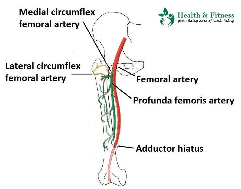
ёяшэ Where Is Your юааfemoralюаб юааarteryюаб What Is юааfemoralюаб юааarteryюаб Dissection The color flow image shows the common femoral artery bifurcation and the location of the pulsed doppler sample volume. the flow pattern in the center stream of normal lower extremity arteries is relatively uniform, with the red blood cells all having nearly the same velocity. Anatomical variations. there are numerous anatomical variations in the lower limb venous system, and even experienced sonographers will encounter new variations from time to time. duplicated, or bifid, vein systems are relatively common and mainly involve the femoral vein and popliteal vein ( fig. 13.9 ). The femoral artery is a large blood vessel that provides oxygenated blood to lower extremity structures and in part to the lower anterior abdominal wall. the common femoral artery arises as a continuation of the external iliac artery after it passes under the inguinal ligament. the femoral artery, vein, and nerve all exist in the anterior region of the thigh known as the femoral triangle, just. The flow of blood in the common femoral, popliteal, dorsalis pedis, and posterior tibial arteries and in any prosthetic or venous bypass grafts can be assessed with the use of doppler.

Information About Femoral Artery Its Branches Personalcarenheal The femoral artery is a large blood vessel that provides oxygenated blood to lower extremity structures and in part to the lower anterior abdominal wall. the common femoral artery arises as a continuation of the external iliac artery after it passes under the inguinal ligament. the femoral artery, vein, and nerve all exist in the anterior region of the thigh known as the femoral triangle, just. The flow of blood in the common femoral, popliteal, dorsalis pedis, and posterior tibial arteries and in any prosthetic or venous bypass grafts can be assessed with the use of doppler.

Comments are closed.