The Anatomy Of The Heart A Crash Course With Dissection

Heart Dissection This lecture outlines the structure and function of heart. specifically, the anatomical position, fibrous skeleton, flow of blood, muscular walls and conduct. Your heart gets a lot of attention from poets, songwriters, and storytellers, but today hank's gonna tell you how it really works. the heart’s ventricles, at.

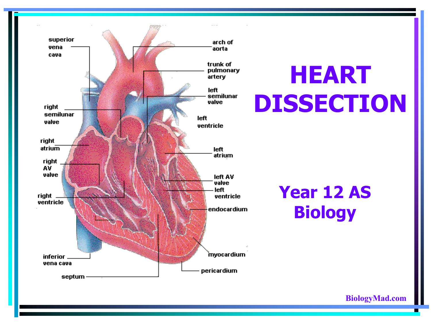
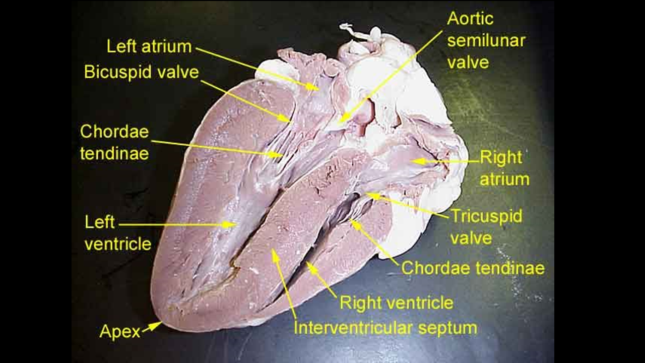
Heart Dissection Photo Gallery Scientist Cindy Anatomy of the heart made easy along with the blood flow through the cardiac structures, valves, atria, and ventricles. cardiovascular system animation for u. The following video is a step by step dissection of the heart and middle mediastinum. even though this dissection has just a few cuts and some cleaning, the anatomy of the heart may be challenging. with this dissection, a little bit of prep will go a long way. category: cardiovascular, human dissection labs, human dissection labs, pre clinical. 3. aorta and the pulmonary arteries. several vessels extend from the superior (topmost) side of the heart. it may be necessary to remove excessive tissue to visualize structures and for ease of dissection. identify the aorta and the pulmonary artery and then cut them to make the valves visible (figure 5.4). Anatomy of the heart: anatomical illustrations and structures, 3d model and photographs of dissection. this interactive atlas of human heart anatomy is based on medical illustrations and cadaver photography. the user can show or hide the anatomical labels which provide a useful tool to create illustrations perfectly adapted for teaching.

Cardiovascular Lab Heart Frontal Dissection Diagram Quizlet 3. aorta and the pulmonary arteries. several vessels extend from the superior (topmost) side of the heart. it may be necessary to remove excessive tissue to visualize structures and for ease of dissection. identify the aorta and the pulmonary artery and then cut them to make the valves visible (figure 5.4). Anatomy of the heart: anatomical illustrations and structures, 3d model and photographs of dissection. this interactive atlas of human heart anatomy is based on medical illustrations and cadaver photography. the user can show or hide the anatomical labels which provide a useful tool to create illustrations perfectly adapted for teaching. Heart anatomy. the heart has five surfaces: base (posterior), diaphragmatic (inferior), sternocostal (anterior), and left and right pulmonary surfaces. it also has several margins: right, left, superior, and inferior: the right margin is the small section of the right atrium that extends between the superior and inferior vena cava . 3. aorta and the pulmonary arteries. several vessels extend from the superior (topmost) side of the heart. it may be necessary to remove excessive tissue to visualize structures and for ease of dissection. identify the aorta and the pulmonary artery and then cut them to make the valves visible (figure 5.4).
/human-heart-circulatory-system-598167278-5c48d4d2c9e77c0001a577d4.jpg)
Av And Semilunar Heart Valves Heart anatomy. the heart has five surfaces: base (posterior), diaphragmatic (inferior), sternocostal (anterior), and left and right pulmonary surfaces. it also has several margins: right, left, superior, and inferior: the right margin is the small section of the right atrium that extends between the superior and inferior vena cava . 3. aorta and the pulmonary arteries. several vessels extend from the superior (topmost) side of the heart. it may be necessary to remove excessive tissue to visualize structures and for ease of dissection. identify the aorta and the pulmonary artery and then cut them to make the valves visible (figure 5.4).

Comments are closed.